 Movie
Movie Controller


+ Open data

- Basic information
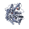
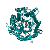
| Entry | Database: PDB / ID: 6m6m | ||||||
|---|---|---|---|---|---|---|---|
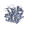
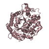
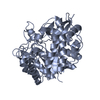
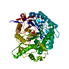
| Title | The crystal structure of glycosidase mutant | ||||||
 Components Components | Beta-glucosidase | ||||||
 Keywords Keywords | HYDROLASE / Glycosidase / Notoginsenoside / Mutant | ||||||
| Function / homology |  Function and homology information Function and homology informationbeta-glucosidase / beta-glucosidase activity / cellulose catabolic process / cytosol Similarity search - Function | ||||||
| Biological species |  Kribbella flavida (bacteria) Kribbella flavida (bacteria) | ||||||
| Method |  X-RAY DIFFRACTION / SYNCHROTRON / MOLECULAR REPLACEMENT / Resolution: 2.07 Å X-RAY DIFFRACTION / SYNCHROTRON / MOLECULAR REPLACEMENT / Resolution: 2.07 Å | ||||||
 Authors Authors | Wang, R.F. | ||||||
 Citation Citation | Journal: To Be Published Title: Characterization and Structural Elucidation of Enhanced Catalytic Activity upon Engineering Glycosidase KfGH01 for the Production of Vina-ginsenoside R7 Authors: Wang, R.F. | ||||||
| History |
|
- Structure visualization
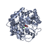
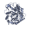
| Structure viewer | Molecule: MolmilJmol/JSmol |
|---|
- Downloads & links
-Download
| PDBx/mmCIF format | 6m6m.cif.gz | 211.7 KB | Display | PDBx/mmCIF format |
|---|---|---|---|---|
| PDB format | pdb6m6m.ent.gz | 165.9 KB | Display | PDB format |
| PDBx/mmJSON format | 6m6m.json.gz | Tree view | PDBx/mmJSON format | |
| Others |  Other downloads Other downloads |
-Validation report
| Arichive directory | https://data.pdbj.org/pub/pdb/validation_reports/m6/6m6mftp://data.pdbj.org/pub/pdb/validation_reports/m6/6m6m | HTTPS FTP |
|---|
-Related structure data
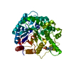
| Related structure data |  6m6lC  3ta9S S: Starting model for refinement C: citing same article ( |
|---|---|
| Similar structure data |

-Links
 PDBj
PDBj- Assembly
| Deposited unit | 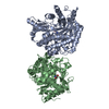
| ||||||||
|---|---|---|---|---|---|---|---|---|---|
| 1 | 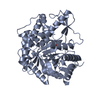
| ||||||||
| 2 | 
| ||||||||
| Unit cell |
|
-Components
| #1: Protein | Mass: 49359.059 Da / Num. of mol.: 2 / Mutation: I248F, Y410R Source method: isolated from a genetically manipulated source Source: (gene. exp.) Kribbella flavida (strain DSM 17836 / JCM 10339 / NBRC 14399) (bacteria)Strain: DSM 17836 / JCM 10339 / NBRC 14399 / Gene: Kfla_5268 Production host: References: UniProt: D2PL27, beta-glucosidase #2: Chemical |   Mass: 122.143 Da / Num. of mol.: 2 / Source method: obtained synthetically / Formula: C4H12NO3 / Feature type: SUBJECT OF INVESTIGATION / Comment: pH buffer*YM Mass: 122.143 Da / Num. of mol.: 2 / Source method: obtained synthetically / Formula: C4H12NO3 / Feature type: SUBJECT OF INVESTIGATION / Comment: pH buffer*YM#3: Water | ChemComp-HOH / |  Mass: 18.015 Da / Num. of mol.: 1195 / Source method: isolated from a natural source / Formula: H2O Mass: 18.015 Da / Num. of mol.: 1195 / Source method: isolated from a natural source / Formula: H2OHas ligand of interest | Y | |
|---|
-Experimental details
-Experiment
| Experiment | Method: X-RAY DIFFRACTION / Number of used crystals: 1 |
|---|
- Sample preparation
| Crystal | Density Matthews: 2.38 Å3/Da / Density % sol: 48.32 % |
|---|---|
| Crystal grow | Temperature: 293 K / Method: vapor diffusion, sitting drop / pH: 8.5 Details: 0.1 M Tris-HCl (pH 8.5), 0.2 M Magnesium chloride, PEG 8000 (20%, w/v) |
-Data collection
| Diffraction | Mean temperature: 100 K / Serial crystal experiment: N |
|---|---|
| Diffraction source | Source: SYNCHROTRON / Site: SSRF  / Beamline: BL17U / Wavelength: 0.9791 Å / Beamline: BL17U / Wavelength: 0.9791 Å |
| Detector | Type: ADSC QUANTUM 315r / Detector: CCD / Date: Oct 28, 2018 |
| Radiation | Protocol: SINGLE WAVELENGTH / Monochromatic (M) / Laue (L): M / Scattering type: x-ray |
| Radiation wavelength | Wavelength: 0.9791 Å / Relative weight: 1 |
| Reflection | Resolution: 2.07→50 Å / Num. obs: 342172 / % possible obs: 99.9 % / Redundancy: 3.7 % / CC1/2: 0.99 / Net I/σ(I): 27.6 |
| Reflection shell | Resolution: 2.07→2.11 Å / Num. unique obs: 342172 / CC1/2: 0.99 |
- Processing
| Software |
| |||||||||||||||||||||||||||||||||||||||||||||||||||||||||||||||||||||||||||||||||||||||||||||||||||||||||
|---|---|---|---|---|---|---|---|---|---|---|---|---|---|---|---|---|---|---|---|---|---|---|---|---|---|---|---|---|---|---|---|---|---|---|---|---|---|---|---|---|---|---|---|---|---|---|---|---|---|---|---|---|---|---|---|---|---|---|---|---|---|---|---|---|---|---|---|---|---|---|---|---|---|---|---|---|---|---|---|---|---|---|---|---|---|---|---|---|---|---|---|---|---|---|---|---|---|---|---|---|---|---|---|---|---|---|
| Refinement | Method to determine structure: MOLECULAR REPLACEMENT Starting model: 3TA9 Resolution: 2.07→32.634 Å / SU ML: 0.14 / Cross valid method: FREE R-VALUE / σ(F): 1.35 / Phase error: 16.39
| |||||||||||||||||||||||||||||||||||||||||||||||||||||||||||||||||||||||||||||||||||||||||||||||||||||||||
| Solvent computation | Shrinkage radii: 0.9 Å / VDW probe radii: 1.11 Å | |||||||||||||||||||||||||||||||||||||||||||||||||||||||||||||||||||||||||||||||||||||||||||||||||||||||||
| Refinement step | Cycle: LAST / Resolution: 2.07→32.634 Å
| |||||||||||||||||||||||||||||||||||||||||||||||||||||||||||||||||||||||||||||||||||||||||||||||||||||||||
| Refine LS restraints |
| |||||||||||||||||||||||||||||||||||||||||||||||||||||||||||||||||||||||||||||||||||||||||||||||||||||||||
| LS refinement shell |
|
