 Movie
Movie Controller

[English] 日本語
 Yorodumi
- PDB-1np2: Crystal structure of thermostable beta-glycosidase from thermophi... -
+ Open data

- Basic information
| Entry | Database: PDB / ID: 1np2 | ||||||
|---|---|---|---|---|---|---|---|
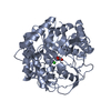
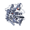
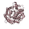
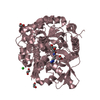
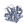
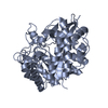
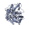
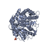
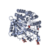
| Title | Crystal structure of thermostable beta-glycosidase from thermophilic eubacterium Thermus nonproteolyticus HG102 | ||||||
 Components Components | beta-glycosidase | ||||||
 Keywords Keywords | HYDROLASE / TIM barrel | ||||||
| Function / homology |  Function and homology information Function and homology informationbeta-glucosidase / beta-glucosidase activity / cellulose catabolic process / cytosol Similarity search - Function | ||||||
| Biological species |   Thermus nonproteolyticus (bacteria) Thermus nonproteolyticus (bacteria) | ||||||
| Method |  X-RAY DIFFRACTION / SYNCHROTRON / MOLECULAR REPLACEMENT / Resolution: 2.4 Å X-RAY DIFFRACTION / SYNCHROTRON / MOLECULAR REPLACEMENT / Resolution: 2.4 Å | ||||||
 Authors Authors | Liang, D.C. / Chang, W.R. / Wang, X.Q. / He, X.Y. | ||||||
 Citation Citation | Journal: J.Bacteriol. / Year: 2003 Title: Structural Basis for Thermostability of beta-Glycosidase from the Thermophilic Eubacterium Thermus nonproteolyticus HG102. Authors: Wang, X. / He, X. / Yang, S. / An, X. / Chang, W. / Liang, D. #1: Journal: Acta Crystallogr.,Sect.D / Year: 2001Title: Overexpression, purification, crystallization and preliminary crystallographic studies on a thermostable beta-glycosidase from Thermus nonproteolyticus HG102. Authors: He, X.Y. / Wang, X.Q. / Yang, S.J. / Chang, W.R. / Liang, D.C. | ||||||
| History |
|
- Structure visualization
| Structure viewer | Molecule: MolmilJmol/JSmol |
|---|
- Downloads & links
-Download
| PDBx/mmCIF format | 1np2.cif.gz | 181.6 KB | Display | PDBx/mmCIF format |
|---|---|---|---|---|
| PDB format | pdb1np2.ent.gz | 145.8 KB | Display | PDB format |
| PDBx/mmJSON format | 1np2.json.gz | Tree view | PDBx/mmJSON format | |
| Others |  Other downloads Other downloads |
-Validation report
| Arichive directory | https://data.pdbj.org/pub/pdb/validation_reports/np/1np2ftp://data.pdbj.org/pub/pdb/validation_reports/np/1np2 | HTTPS FTP |
|---|
-Related structure data
| Related structure data |  1bgaS S: Starting model for refinement |
|---|---|
| Similar structure data |

-Links
 PDBj
PDBj- Assembly
| Deposited unit | 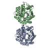
| ||||||||
|---|---|---|---|---|---|---|---|---|---|
| 1 | 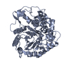
| ||||||||
| 2 | 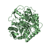
| ||||||||
| Unit cell |
| ||||||||
| Details | monomer enzyme |
-Components
| #1: Protein | Mass: 49056.168 Da / Num. of mol.: 2 Source method: isolated from a genetically manipulated source Source: (gene. exp.) Thermus nonproteolyticus (bacteria) / Strain: HG102 / Plasmid: pET21a / Species (production host): Escherichia coli / Production host: #2: Water | ChemComp-HOH / |  Mass: 18.015 Da / Num. of mol.: 334 / Source method: isolated from a natural source / Formula: H2O Mass: 18.015 Da / Num. of mol.: 334 / Source method: isolated from a natural source / Formula: H2O |
|---|
-Experimental details
-Experiment
| Experiment | Method: X-RAY DIFFRACTION / Number of used crystals: 1 |
|---|
- Sample preparation
| Crystal | Density Matthews: 2.79 Å3/Da / Density % sol: 55.52 % | ||||||||||||||||||||||||||||||||||||||||||
|---|---|---|---|---|---|---|---|---|---|---|---|---|---|---|---|---|---|---|---|---|---|---|---|---|---|---|---|---|---|---|---|---|---|---|---|---|---|---|---|---|---|---|---|
| Crystal grow | Temperature: 291 K / Method: vapor diffusion, hanging drop / pH: 4.6 Details: MPD, pH 4.6, VAPOR DIFFUSION, HANGING DROP, temperature 291K | ||||||||||||||||||||||||||||||||||||||||||
| Crystal grow | *PLUS Temperature: 291 K / pH: 7.5 / Method: vapor diffusion, hanging dropDetails: He, X.Y., (2001) Acta Crystallogr.,Sect.D, 57, 1650. | ||||||||||||||||||||||||||||||||||||||||||
| Components of the solutions | *PLUS
|
-Data collection
| Diffraction | Mean temperature: 100 K |
|---|---|
| Diffraction source | Source: SYNCHROTRON / Site: Photon Factory  / Beamline: BL-6B / Wavelength: 1 Å / Beamline: BL-6B / Wavelength: 1 Å |
| Detector | Type: WEISSENBERG / Detector: DIFFRACTOMETER / Date: Dec 3, 2001 |
| Radiation | Protocol: SINGLE WAVELENGTH / Monochromatic (M) / Laue (L): M / Scattering type: x-ray |
| Radiation wavelength | Wavelength: 1 Å / Relative weight: 1 |
| Reflection | Resolution: 2.4→83.51 Å / Num. all: 44647 / Num. obs: 42023 / % possible obs: 94 % / Observed criterion σ(F): 0 / Observed criterion σ(I): -3 / Rmerge(I) obs: 0.182 / Net I/σ(I): 9.9 |
| Reflection shell | Resolution: 2.4→2.49 Å / Rmerge(I) obs: 0.328 / Mean I/σ(I) obs: 2.2 / % possible all: 73.4 |
| Reflection | *PLUS Lowest resolution: 20 Å / Num. obs: 41963 / % possible obs: 94 % |
| Reflection shell | *PLUS % possible obs: 73.4 % / Rmerge(I) obs: 0.327 |
- Processing
| Software |
| |||||||||||||||||||||||||
|---|---|---|---|---|---|---|---|---|---|---|---|---|---|---|---|---|---|---|---|---|---|---|---|---|---|---|
| Refinement | Method to determine structure: MOLECULAR REPLACEMENT Starting model: PDB code 1BGA Resolution: 2.4→50 Å / σ(F): 0 / Stereochemistry target values: Engh & Huber
| |||||||||||||||||||||||||
| Refine analyze | Luzzati coordinate error obs: 0.35 Å / Luzzati d res low obs: 5 Å / Luzzati sigma a obs: 0.34 Å | |||||||||||||||||||||||||
| Refinement step | Cycle: LAST / Resolution: 2.4→50 Å
| |||||||||||||||||||||||||
| Refine LS restraints |
| |||||||||||||||||||||||||
| Refinement | *PLUS Lowest resolution: 20 Å / Rfactor Rwork: 0.23 | |||||||||||||||||||||||||
| Solvent computation | *PLUS | |||||||||||||||||||||||||
| Displacement parameters | *PLUS | |||||||||||||||||||||||||
| Refine LS restraints | *PLUS
|
