 Movie
Movie Controller

[English] 日本語
 Yorodumi
- PDB-6m55: Crystal structure of the E496A mutant of HsBglA in complex with 4... -
+ Open data

- Basic information
| Entry | Database: PDB / ID: 6m55 | |||||||||
|---|---|---|---|---|---|---|---|---|---|---|
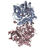
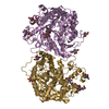
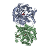
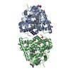
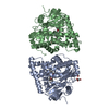
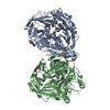
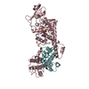
| Title | Crystal structure of the E496A mutant of HsBglA in complex with 4-galactosyllactose | |||||||||
 Components Components | Beta-galactosidase-like enzyme | |||||||||
 Keywords Keywords | HYDROLASE / galactooligosaccharide / transgalactosylation / beta-galactosidase / beta-glucosidase / Hamamotoa singularis / STRUCTURAL PROTEIN | |||||||||
| Function / homology |  Function and homology information Function and homology informationbeta-glucosidase activity / carbohydrate metabolic process / metal ion binding Similarity search - Function | |||||||||
| Biological species |  Hamamotoa singularis (fungus) Hamamotoa singularis (fungus) | |||||||||
| Method |  X-RAY DIFFRACTION / SYNCHROTRON / MOLECULAR REPLACEMENT / Resolution: 3 Å X-RAY DIFFRACTION / SYNCHROTRON / MOLECULAR REPLACEMENT / Resolution: 3 Å | |||||||||
 Authors Authors | Uehara, R. / Iwamoto, R. / Aoki, S. / Yoshizawa, T. / Takano, K. / Matsumura, H. / Tanaka, S.-i. | |||||||||
| Funding support |  Japan, 2items Japan, 2items
| |||||||||
 Citation Citation | Journal: Protein Sci. / Year: 2020 Title: Crystal structure of a GH1 beta-glucosidase from Hamamotoa singularis. Authors: Uehara, R. / Iwamoto, R. / Aoki, S. / Yoshizawa, T. / Takano, K. / Matsumura, H. / Tanaka, S.I. | |||||||||
| History |
|
- Structure visualization
| Structure viewer | Molecule: MolmilJmol/JSmol |
|---|
- Downloads & links
-Download
| PDBx/mmCIF format | 6m55.cif.gz | 202.7 KB | Display | PDBx/mmCIF format |
|---|---|---|---|---|
| PDB format | pdb6m55.ent.gz | 160.9 KB | Display | PDB format |
| PDBx/mmJSON format | 6m55.json.gz | Tree view | PDBx/mmJSON format | |
| Others |  Other downloads Other downloads |
-Validation report
| Arichive directory | https://data.pdbj.org/pub/pdb/validation_reports/m5/6m55ftp://data.pdbj.org/pub/pdb/validation_reports/m5/6m55 | HTTPS FTP |
|---|
-Related structure data
| Related structure data |  6m4eC  6m4fC  4gxpS S: Starting model for refinement C: citing same article ( |
|---|---|
| Similar structure data |


-Links
 PDBj
PDBj


- Assembly
| Deposited unit | 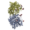
| ||||||||
|---|---|---|---|---|---|---|---|---|---|
| 1 |
| ||||||||
| Unit cell |
|
-Components
-Protein / Non-polymers , 2 types, 5 molecules AD
| #1: Protein | Mass: 60452.949 Da / Num. of mol.: 2 / Mutation: E496A Source method: isolated from a genetically manipulated source Source: (gene. exp.) Hamamotoa singularis (fungus) / Gene: bglA / Plasmid: pPICZalphaA / Production host: Komagataella pastoris (fungus) / Strain (production host): X-33 / References: UniProt: Q564N5#6: Water | ChemComp-HOH / | Mass: 18.015 Da / Num. of mol.: 3 / Source method: isolated from a natural source / Formula: H2O |
|---|
-Sugars , 4 types, 10 molecules 

| #2: Polysaccharide | Source method: isolated from a genetically manipulated source #3: Polysaccharide | Source method: isolated from a genetically manipulated source #4: Sugar |  Type: D-saccharide, alpha linking / Mass: 180.156 Da / Num. of mol.: 2 Type: D-saccharide, alpha linking / Mass: 180.156 Da / Num. of mol.: 2Source method: isolated from a genetically manipulated source Formula: C6H12O6 #5: Sugar | ChemComp-NAG /  Type: D-saccharide, beta linking / Mass: 221.208 Da / Num. of mol.: 4 Type: D-saccharide, beta linking / Mass: 221.208 Da / Num. of mol.: 4Source method: isolated from a genetically manipulated source Formula: C8H15NO6 |
|---|
-Details
| Has ligand of interest | Y |
|---|---|
| Has protein modification | Y |
-Experimental details
-Experiment
| Experiment | Method: X-RAY DIFFRACTION / Number of used crystals: 1 |
|---|
- Sample preparation
| Crystal | Density Matthews: 2.22 Å3/Da / Density % sol: 44.51 % |
|---|---|
| Crystal grow | Temperature: 293 K / Method: vapor diffusion, hanging drop / pH: 7 / Details: 0.1 M SPG Buffer pH 7.0, 27% (w/v) PEG 1500 |
-Data collection
| Diffraction | Mean temperature: 100 K / Serial crystal experiment: N |
|---|---|
| Diffraction source | Source: SYNCHROTRON / Site: SPring-8 / Beamline: BL44XU / Wavelength: 0.9 Å |
| Detector | Type: DECTRIS EIGER X 16M / Detector: PIXEL / Date: Nov 23, 2019 |
| Radiation | Protocol: SINGLE WAVELENGTH / Monochromatic (M) / Laue (L): M / Scattering type: x-ray |
| Radiation wavelength | Wavelength: 0.9 Å / Relative weight: 1 |
| Reflection | Resolution: 2.9→45.99 Å / Num. obs: 24658 / % possible obs: 96.66 % / Redundancy: 6.1 % / Biso Wilson estimate: 57.01 Å2 / CC1/2: 0.993 / Rmerge(I) obs: 0.1612 / Rpim(I) all: 0.06944 / Rrim(I) all: 0.1759 / Net I/σ(I): 7.27 |
| Reflection shell | Resolution: 2.9→2.98 Å / Redundancy: 4.9 % / Rmerge(I) obs: 0.5642 / Mean I/σ(I) obs: 1.57 / Num. unique obs: 1795 / CC1/2: 0.912 / Rpim(I) all: 0.2658 / Rrim(I) all: 0.6255 / % possible all: 96.2 |
- Processing
| Software |
| |||||||||||||||||||||||||||||||||||||||||||||||||||||||||||||||
|---|---|---|---|---|---|---|---|---|---|---|---|---|---|---|---|---|---|---|---|---|---|---|---|---|---|---|---|---|---|---|---|---|---|---|---|---|---|---|---|---|---|---|---|---|---|---|---|---|---|---|---|---|---|---|---|---|---|---|---|---|---|---|---|---|
| Refinement | Method to determine structure: MOLECULAR REPLACEMENT Starting model: 4GXP Resolution: 3→45.21 Å / SU ML: 0.48 / Cross valid method: THROUGHOUT / σ(F): 1.37 / Phase error: 34.02 / Stereochemistry target values: ML
| |||||||||||||||||||||||||||||||||||||||||||||||||||||||||||||||
| Solvent computation | Shrinkage radii: 0.9 Å / VDW probe radii: 1.11 Å / Solvent model: FLAT BULK SOLVENT MODEL | |||||||||||||||||||||||||||||||||||||||||||||||||||||||||||||||
| Displacement parameters | Biso max: 57.8 Å2 / Biso mean: 55.3936 Å2 / Biso min: 20 Å2 | |||||||||||||||||||||||||||||||||||||||||||||||||||||||||||||||
| Refinement step | Cycle: final / Resolution: 3→45.21 Å
| |||||||||||||||||||||||||||||||||||||||||||||||||||||||||||||||
| LS refinement shell | Refine-ID: X-RAY DIFFRACTION / Rfactor Rfree error: 0 / Total num. of bins used: 8
|
