+ Open data
- Basic information
| Entry | Database: PDB / ID: 6m3b | ||||||
|---|---|---|---|---|---|---|---|
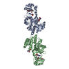
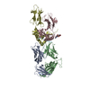
| Title | hAPC-c25k23 Fab complex | ||||||
 Components Components |
| ||||||
 Keywords Keywords | BLOOD CLOTTING/IMMUNE SYSTEM / human APC / c25k23 Fab / complex / BLOOD CLOTTING / BLOOD CLOTTING-IMMUNE SYSTEM complex | ||||||
| Function / homology |  Function and homology information Function and homology informationactivated protein C (thrombin-activated peptidase) / positive regulation of establishment of endothelial barrier / negative regulation of coagulation / negative regulation of blood coagulation / Transport of gamma-carboxylated protein precursors from the endoplasmic reticulum to the Golgi apparatus / Gamma-carboxylation of protein precursors / Common Pathway of Fibrin Clot Formation / Removal of aminoterminal propeptides from gamma-carboxylated proteins / Intrinsic Pathway of Fibrin Clot Formation / Cell surface interactions at the vascular wall ...activated protein C (thrombin-activated peptidase) / positive regulation of establishment of endothelial barrier / negative regulation of coagulation / negative regulation of blood coagulation / Transport of gamma-carboxylated protein precursors from the endoplasmic reticulum to the Golgi apparatus / Gamma-carboxylation of protein precursors / Common Pathway of Fibrin Clot Formation / Removal of aminoterminal propeptides from gamma-carboxylated proteins / Intrinsic Pathway of Fibrin Clot Formation / Cell surface interactions at the vascular wall / Post-translational protein phosphorylation / Golgi lumen / negative regulation of inflammatory response / Regulation of Insulin-like Growth Factor (IGF) transport and uptake by Insulin-like Growth Factor Binding Proteins (IGFBPs) / blood coagulation / endoplasmic reticulum lumen / serine-type endopeptidase activity / calcium ion binding / negative regulation of apoptotic process / endoplasmic reticulum / Golgi apparatus / proteolysis / extracellular space / extracellular region Similarity search - Function | ||||||
| Biological species |  Homo sapiens (human) Homo sapiens (human) | ||||||
| Method |  X-RAY DIFFRACTION / X-RAY DIFFRACTION /  SYNCHROTRON / SYNCHROTRON /  MOLECULAR REPLACEMENT / Resolution: 2.2 Å MOLECULAR REPLACEMENT / Resolution: 2.2 Å | ||||||
 Authors Authors | Wang, X. / Li, L. / Zhao, X. / Egner, U. | ||||||
 Citation Citation |  Journal: Nat Commun / Year: 2020 Journal: Nat Commun / Year: 2020Title: Targeted inhibition of activated protein C by a non-active-site inhibitory antibody to treat hemophilia. Authors: Zhao, X.Y. / Wilmen, A. / Wang, D. / Wang, X. / Bauzon, M. / Kim, J.Y. / Linden, L. / Li, L. / Egner, U. / Marquardt, T. / Moosmayer, D. / Tebbe, J. / Gluck, J.M. / Ellinger, P. / McLean, K. ...Authors: Zhao, X.Y. / Wilmen, A. / Wang, D. / Wang, X. / Bauzon, M. / Kim, J.Y. / Linden, L. / Li, L. / Egner, U. / Marquardt, T. / Moosmayer, D. / Tebbe, J. / Gluck, J.M. / Ellinger, P. / McLean, K. / Yuan, S. / Yegneswaran, S. / Jiang, X. / Evans, V. / Gu, J.M. / Schneider, D. / Zhu, Y. / Xu, Y. / Mallari, C. / Hesslein, A. / Wang, Y. / Schmidt, N. / Gutberlet, K. / Ruehl-Fehlert, C. / Freyberger, A. / Hermiston, T. / Patel, C. / Sim, D. / Mosnier, L.O. / Laux, V. | ||||||
| History |
|
- Structure visualization
| Structure viewer | Molecule:  Molmil Molmil Jmol/JSmol Jmol/JSmol |
|---|
- Downloads & links
- Download
| PDBx/mmCIF format |  6m3b.cif.gz 6m3b.cif.gz | 165.3 KB | Display |  PDBx/mmCIF format PDBx/mmCIF format |
|---|---|---|---|---|
| PDB format |  pdb6m3b.ent.gz pdb6m3b.ent.gz | 125.6 KB | Display |  PDB format PDB format |
| PDBx/mmJSON format |  6m3b.json.gz 6m3b.json.gz | Tree view |  PDBx/mmJSON format PDBx/mmJSON format | |
| Others |  Other downloads Other downloads |
-Validation report
| Summary document |  6m3b_validation.pdf.gz 6m3b_validation.pdf.gz | 478.4 KB | Display |  wwPDB validaton report wwPDB validaton report |
|---|---|---|---|---|
| Full document |  6m3b_full_validation.pdf.gz 6m3b_full_validation.pdf.gz | 489.4 KB | Display | |
| Data in XML |  6m3b_validation.xml.gz 6m3b_validation.xml.gz | 29.4 KB | Display | |
| Data in CIF |  6m3b_validation.cif.gz 6m3b_validation.cif.gz | 40.9 KB | Display | |
| Arichive directory |  https://data.pdbj.org/pub/pdb/validation_reports/m3/6m3b https://data.pdbj.org/pub/pdb/validation_reports/m3/6m3b ftp://data.pdbj.org/pub/pdb/validation_reports/m3/6m3b ftp://data.pdbj.org/pub/pdb/validation_reports/m3/6m3b | HTTPS FTP |
-Related structure data
| Related structure data |  6m3cC 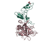 1autS S: Starting model for refinement C: citing same article ( |
|---|---|
| Similar structure data |
- Links
Links
- Assembly
| Deposited unit | 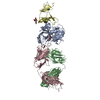
| ||||||||
|---|---|---|---|---|---|---|---|---|---|
| 1 |
| ||||||||
| Unit cell |
|
- Components
-Vitamin K-dependent protein C ... , 2 types, 2 molecules AD
| #1: Protein | Mass: 28091.115 Da / Num. of mol.: 1 Source method: isolated from a genetically manipulated source Source: (gene. exp.)  Homo sapiens (human) / Gene: PROC / Production host: Homo sapiens (human) / Gene: PROC / Production host:  Homo sapiens (human) Homo sapiens (human)References: UniProt: P04070, activated protein C (thrombin-activated peptidase) |
|---|---|
| #2: Protein | Mass: 17607.980 Da / Num. of mol.: 1 Source method: isolated from a genetically manipulated source Source: (gene. exp.)  Homo sapiens (human) / Gene: PROC / Production host: Homo sapiens (human) / Gene: PROC / Production host:  Homo sapiens (human) Homo sapiens (human)References: UniProt: P04070, activated protein C (thrombin-activated peptidase) |
-Antibody , 2 types, 2 molecules BC
| #3: Antibody | Mass: 22472.770 Da / Num. of mol.: 1 Source method: isolated from a genetically manipulated source Source: (gene. exp.)  Homo sapiens (human) / Production host: Homo sapiens (human) / Production host:  |
|---|---|
| #4: Antibody | Mass: 23890.752 Da / Num. of mol.: 1 Source method: isolated from a genetically manipulated source Source: (gene. exp.)  Homo sapiens (human) / Production host: Homo sapiens (human) / Production host:  |
-Sugars / Non-polymers , 2 types, 141 molecules 


| #5: Sugar | | #6: Water | ChemComp-HOH / | |
|---|
-Details
| Has ligand of interest | N |
|---|---|
| Has protein modification | Y |
-Experimental details
-Experiment
| Experiment | Method:  X-RAY DIFFRACTION / Number of used crystals: 1 X-RAY DIFFRACTION / Number of used crystals: 1 |
|---|
- Sample preparation
| Crystal | Density Matthews: 3.19 Å3/Da / Density % sol: 61.41 % |
|---|---|
| Crystal grow | Temperature: 291 K / Method: vapor diffusion, sitting drop Details: 0.1% n-Octyl-beta-D-glucoside, 0.1M sodium citrate tribasic dehydrate pH 5.5, 22% (w/v) PEG 3350 |
-Data collection
| Diffraction | Mean temperature: 100 K / Serial crystal experiment: N |
|---|---|
| Diffraction source | Source:  SYNCHROTRON / Site: SYNCHROTRON / Site:  SSRF SSRF  / Beamline: BL17U1 / Wavelength: 1 Å / Beamline: BL17U1 / Wavelength: 1 Å |
| Detector | Type: ADSC QUANTUM 315r / Detector: CCD / Date: Apr 21, 2012 |
| Radiation | Protocol: SINGLE WAVELENGTH / Monochromatic (M) / Laue (L): M / Scattering type: x-ray |
| Radiation wavelength | Wavelength: 1 Å / Relative weight: 1 |
| Reflection | Resolution: 2.2→30 Å / Num. obs: 57099 / % possible obs: 97.22 % / Redundancy: 4.2 % / CC1/2: 0.991 / Rmerge(I) obs: 0.096 / Net I/σ(I): 32.22 |
| Reflection shell | Resolution: 2.2→2.26 Å / Rmerge(I) obs: 0.989 / Mean I/σ(I) obs: 3.04 / Num. unique obs: 4288 / CC1/2: 0.825 / Rpim(I) all: 0.531 |
- Processing
| Software |
| ||||||||||||||||||||||||||||||||||||||||||||||||||||||||||||||||||||||||||||||||||||||||||||||||||||||||||||||||||||||||||||||||||||
|---|---|---|---|---|---|---|---|---|---|---|---|---|---|---|---|---|---|---|---|---|---|---|---|---|---|---|---|---|---|---|---|---|---|---|---|---|---|---|---|---|---|---|---|---|---|---|---|---|---|---|---|---|---|---|---|---|---|---|---|---|---|---|---|---|---|---|---|---|---|---|---|---|---|---|---|---|---|---|---|---|---|---|---|---|---|---|---|---|---|---|---|---|---|---|---|---|---|---|---|---|---|---|---|---|---|---|---|---|---|---|---|---|---|---|---|---|---|---|---|---|---|---|---|---|---|---|---|---|---|---|---|---|---|
| Refinement | Method to determine structure:  MOLECULAR REPLACEMENT MOLECULAR REPLACEMENTStarting model: 1AUT Resolution: 2.2→27.542 Å / SU ML: 0.31 / Cross valid method: THROUGHOUT / σ(F): 1.34 / Phase error: 33.84
| ||||||||||||||||||||||||||||||||||||||||||||||||||||||||||||||||||||||||||||||||||||||||||||||||||||||||||||||||||||||||||||||||||||
| Solvent computation | Shrinkage radii: 0.9 Å / VDW probe radii: 1.11 Å | ||||||||||||||||||||||||||||||||||||||||||||||||||||||||||||||||||||||||||||||||||||||||||||||||||||||||||||||||||||||||||||||||||||
| Displacement parameters | Biso max: 196.09 Å2 / Biso mean: 65.4415 Å2 / Biso min: 38.39 Å2 | ||||||||||||||||||||||||||||||||||||||||||||||||||||||||||||||||||||||||||||||||||||||||||||||||||||||||||||||||||||||||||||||||||||
| Refinement step | Cycle: final / Resolution: 2.2→27.542 Å
| ||||||||||||||||||||||||||||||||||||||||||||||||||||||||||||||||||||||||||||||||||||||||||||||||||||||||||||||||||||||||||||||||||||
| LS refinement shell | Refine-ID: X-RAY DIFFRACTION / Rfactor Rfree error: 0
|
 Movie
Movie Controller




 PDBj
PDBj


