 Movie
Movie Controller


+ Open data

- Basic information
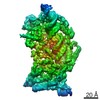
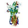
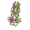
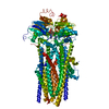
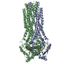
| Entry | Database: PDB / ID: 6lxe | |||||||||||||||||||||
|---|---|---|---|---|---|---|---|---|---|---|---|---|---|---|---|---|---|---|---|---|---|---|
| Title | DROSHA-DGCR8 complex | |||||||||||||||||||||
 Components Components |
| |||||||||||||||||||||
 Keywords Keywords | HYDROLASE/RNA BINDING PROTEIN / Ribonuclease / RNA BINDING PROTEIN / HYDROLASE-RNA BINDING PROTEIN complex | |||||||||||||||||||||
| Function / homology |  Function and homology information Function and homology informationpositive regulation of pre-miRNA processing / microprocessor complex / DEAD/H-box RNA helicase binding / primary miRNA binding / ribonuclease III / protein-RNA adaptor activity / Transcriptional Regulation by MECP2 / primary miRNA processing / ribonuclease III activity / pre-miRNA processing ...positive regulation of pre-miRNA processing / microprocessor complex / DEAD/H-box RNA helicase binding / primary miRNA binding / ribonuclease III / protein-RNA adaptor activity / Transcriptional Regulation by MECP2 / primary miRNA processing / ribonuclease III activity / pre-miRNA processing / MicroRNA (miRNA) biogenesis / SMAD binding / R-SMAD binding / lipopolysaccharide binding / rRNA processing / double-stranded RNA binding / site of double-strand break / defense response to Gram-negative bacterium / nuclear body / protein-macromolecule adaptor activity / defense response to Gram-positive bacterium / postsynaptic density / heme binding / positive regulation of gene expression / DNA damage response / nucleolus / glutamatergic synapse / protein homodimerization activity / RNA binding / nucleoplasm / metal ion binding / identical protein binding / nucleus / cytoplasm Similarity search - Function | |||||||||||||||||||||
| Biological species |  Homo sapiens (human) Homo sapiens (human) | |||||||||||||||||||||
| Method | ELECTRON MICROSCOPY / single particle reconstruction / cryo EM / Resolution: 4.2 Å | |||||||||||||||||||||
 Authors Authors | Jin, W. / Wang, J. / Liu, C.P. / Wang, H.W. / Xu, R.M. | |||||||||||||||||||||
| Funding support |  China, 6items China, 6items
| |||||||||||||||||||||
 Citation Citation | Journal: Mol Cell / Year: 2020 Title: Structural Basis for pri-miRNA Recognition by Drosha. Authors: Wenxing Jin / Jia Wang / Chao-Pei Liu / Hong-Wei Wang / Rui-Ming Xu / Abstract: A commencing and critical step in miRNA biogenesis involves processing of pri-miRNAs in the nucleus by Microprocessor. An important, but not completely understood, question is how Drosha, the ...A commencing and critical step in miRNA biogenesis involves processing of pri-miRNAs in the nucleus by Microprocessor. An important, but not completely understood, question is how Drosha, the catalytic subunit of Microprocessor, binds pri-miRNAs and correctly specifies cleavage sites. Here we report the cryoelectron microscopy structures of the Drosha-DGCR8 complex with and without a pri-miRNA. The RNA-bound structure provides direct visualization of the tertiary structure of pri-miRNA and shows that a helix hairpin in the extended PAZ domain and the mobile basic (MB) helix in the RNase IIIa domain of Drosha coordinate to recognize the single-stranded to double-stranded junction of RNA, whereas the dsRNA binding domain makes extensive contacts with the RNA stem. Furthermore, the RNA-free structure reveals an autoinhibitory conformation of the PAZ helix hairpin. These findings provide mechanistic insights into pri-miRNA cleavage site selection and conformational dynamics governing pri-miRNA recognition by the catalytic component of Microprocessor. | |||||||||||||||||||||
| History |
|
- Structure visualization
| Movie |
Movie viewer |
|---|---|
| Structure viewer | Molecule: MolmilJmol/JSmol |
- Downloads & links
-Download
| PDBx/mmCIF format | 6lxe.cif.gz | 233.2 KB | Display | PDBx/mmCIF format |
|---|---|---|---|---|
| PDB format | pdb6lxe.ent.gz | 156.8 KB | Display | PDB format |
| PDBx/mmJSON format | 6lxe.json.gz | Tree view | PDBx/mmJSON format | |
| Others |  Other downloads Other downloads |
-Validation report
| Arichive directory | https://data.pdbj.org/pub/pdb/validation_reports/lx/6lxeftp://data.pdbj.org/pub/pdb/validation_reports/lx/6lxe | HTTPS FTP |
|---|
-Related structure data
| Related structure data |  30006MC  6lxdC M: map data used to model this data C: citing same article ( |
|---|---|
| Similar structure data |


-Links
 PDBj
PDBj


- Assembly
| Deposited unit | 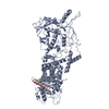
|
|---|---|
| 1 |
|
-Components
| #1: Protein | Mass: 115390.578 Da / Num. of mol.: 1 / Mutation: E1045Q, E1222Q Source method: isolated from a genetically manipulated source Source: (gene. exp.) Homo sapiens (human) / Gene: DROSHA, RN3, RNASE3L, RNASEN / Production host:   Spodoptera frugiperda (fall armyworm) / References: UniProt: Q9NRR4, ribonuclease III Spodoptera frugiperda (fall armyworm) / References: UniProt: Q9NRR4, ribonuclease III | ||||
|---|---|---|---|---|---|
| #2: Protein | Mass: 86171.203 Da / Num. of mol.: 2 Source method: isolated from a genetically manipulated source Source: (gene. exp.) Homo sapiens (human) / Gene: DGCR8, C22orf12, DGCRK6, LP4941 / Production host: Spodoptera frugiperda (fall armyworm) / References: UniProt: Q8WYQ5#3: Chemical |   Mass: 65.409 Da / Num. of mol.: 2 / Source method: obtained synthetically / Formula: Zn Mass: 65.409 Da / Num. of mol.: 2 / Source method: obtained synthetically / Formula: ZnHas ligand of interest | N | |
-Experimental details
-Experiment
| Experiment | Method: ELECTRON MICROSCOPY |
|---|---|
| EM experiment | Aggregation state: PARTICLE / 3D reconstruction method: single particle reconstruction |
- Sample preparation
| Component | Name: Ternary complex of DROSHA-DGCR8 / Type: COMPLEX / Entity ID: #1-#2 / Source: RECOMBINANT |
|---|---|
| Molecular weight | Units: KILODALTONS/NANOMETER / Experimental value: NO |
| Source (natural) | Organism: Homo sapiens (human) |
| Source (recombinant) | Organism: Spodoptera frugiperda (fall armyworm) |
| Buffer solution | pH: 8 |
| Specimen | Conc.: 0.3 mg/ml / Embedding applied: NO / Shadowing applied: NO / Staining applied: NO / Vitrification applied: YES |
| Vitrification | Instrument: FEI VITROBOT MARK IV / Cryogen name: ETHANE / Humidity: 100 % |
- Electron microscopy imaging
| Experimental equipment |  Model: Titan Krios / Image courtesy: FEI Company |
|---|---|
| Microscopy | Model: FEI TITAN KRIOS |
| Electron gun | Electron source:  FIELD EMISSION GUN / Accelerating voltage: 300 kV / Illumination mode: FLOOD BEAM FIELD EMISSION GUN / Accelerating voltage: 300 kV / Illumination mode: FLOOD BEAM |
| Electron lens | Mode: BRIGHT FIELD / Cs: 2.7 mm / C2 aperture diameter: 50 µm |
| Image recording | Electron dose: 50 e/Å2 / Film or detector model: GATAN K2 SUMMIT (4k x 4k) |
- Processing
| Software | Name: PHENIX / Version: 1.16_3549: / Classification: refinement | ||||||||||||||||||||||||
|---|---|---|---|---|---|---|---|---|---|---|---|---|---|---|---|---|---|---|---|---|---|---|---|---|---|
| EM software |
| ||||||||||||||||||||||||
| CTF correction | Type: PHASE FLIPPING AND AMPLITUDE CORRECTION | ||||||||||||||||||||||||
| Symmetry | Point symmetry: C1 (asymmetric) | ||||||||||||||||||||||||
| 3D reconstruction | Resolution: 4.2 Å / Resolution method: FSC 0.143 CUT-OFF / Num. of particles: 109333 / Num. of class averages: 1 / Symmetry type: POINT | ||||||||||||||||||||||||
| Atomic model building | Protocol: RIGID BODY FIT | ||||||||||||||||||||||||
| Atomic model building | PDB-ID: 5B16 Accession code: 5B16 / Source name: PDB / Type: experimental model | ||||||||||||||||||||||||
| Refine LS restraints |
|

