 Movie
Movie Controller

[English] 日本語
 Yorodumi
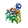
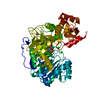
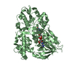
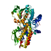
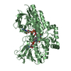
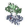
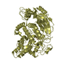
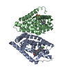
- PDB-5odo: Crystal Structure of the Oleate hydratase of Rhodococcus erythropolis -
+ Open data

- Basic information
| Entry | Database: PDB / ID: 5odo | ||||||
|---|---|---|---|---|---|---|---|
| Title | Crystal Structure of the Oleate hydratase of Rhodococcus erythropolis | ||||||
 Components Components | Isomerase | ||||||
 Keywords Keywords | LYASE / Hydratase / Rossmann fold / FAD binding / fatty acid / immune system | ||||||
| Function / homology | oleate hydratase activity / Oleate hydratase / MCRA family / isomerase activity / FAD binding / fatty acid metabolic process / FAD/NAD(P)-binding domain superfamily / FORMIC ACID / Isomerase Function and homology information Function and homology information | ||||||
| Biological species |  Rhodococcus erythropolis DN1 (bacteria) Rhodococcus erythropolis DN1 (bacteria) | ||||||
| Method |  X-RAY DIFFRACTION / SYNCHROTRON / MOLECULAR REPLACEMENT / Resolution: 2.64 Å X-RAY DIFFRACTION / SYNCHROTRON / MOLECULAR REPLACEMENT / Resolution: 2.64 Å | ||||||
 Authors Authors | Driller, R. / Lorenzen, J. / Waldow, A. / Qoura, F. / Brueck, T. / Loll, B. | ||||||
 Citation Citation | Journal: Chemcatchem / Year: 2017 Title: Rhodococcus erythropolis Oleate Hydratase: a New Member in the Oleate Hydratase Family Tree - Biochemical and Structural Studies. Authors: Lorenzen, J. / Driller, R. / Waldow, A. / Quora, F. / Loll, B. / Brueck, T. | ||||||
| History |
|
- Structure visualization
| Structure viewer | Molecule: MolmilJmol/JSmol |
|---|
- Downloads & links
-Download
| PDBx/mmCIF format | 5odo.cif.gz | 233.5 KB | Display | PDBx/mmCIF format |
|---|---|---|---|---|
| PDB format | pdb5odo.ent.gz | 186.2 KB | Display | PDB format |
| PDBx/mmJSON format | 5odo.json.gz | Tree view | PDBx/mmJSON format | |
| Others |  Other downloads Other downloads |
-Validation report
| Arichive directory | https://data.pdbj.org/pub/pdb/validation_reports/od/5odoftp://data.pdbj.org/pub/pdb/validation_reports/od/5odo | HTTPS FTP |
|---|
-Related structure data
| Related structure data |  4ia5S S: Starting model for refinement |
|---|---|
| Similar structure data |

-Links
 PDBj
PDBj- Assembly
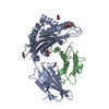
| Deposited unit | 
| ||||||||
|---|---|---|---|---|---|---|---|---|---|
| 1 | 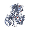
| ||||||||
| 2 | 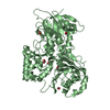
| ||||||||
| Unit cell |
|
-Components
| #1: Protein | Mass: 66031.094 Da / Num. of mol.: 2 Source method: isolated from a genetically manipulated source Source: (gene. exp.) Rhodococcus erythropolis DN1 (bacteria)Gene: N601_10635 / Production host: #2: Chemical |   Mass: 92.094 Da / Num. of mol.: 2 / Source method: obtained synthetically / Formula: C3H8O3 Mass: 92.094 Da / Num. of mol.: 2 / Source method: obtained synthetically / Formula: C3H8O3#3: Chemical | ChemComp-FMT /   Mass: 46.025 Da / Num. of mol.: 14 / Source method: obtained synthetically / Formula: CH2O2 Mass: 46.025 Da / Num. of mol.: 14 / Source method: obtained synthetically / Formula: CH2O2#4: Chemical |   Mass: 24.305 Da / Num. of mol.: 2 / Source method: obtained synthetically / Formula: Mg Mass: 24.305 Da / Num. of mol.: 2 / Source method: obtained synthetically / Formula: Mg#5: Water | ChemComp-HOH / |  Mass: 18.015 Da / Num. of mol.: 429 / Source method: isolated from a natural source / Formula: H2O Mass: 18.015 Da / Num. of mol.: 429 / Source method: isolated from a natural source / Formula: H2O |
|---|
-Experimental details
-Experiment
| Experiment | Method: X-RAY DIFFRACTION / Number of used crystals: 1 |
|---|
- Sample preparation
| Crystal | Density Matthews: 4.77 Å3/Da / Density % sol: 74.19 % |
|---|---|
| Crystal grow | Temperature: 291.15 K / Method: vapor diffusion, hanging drop Details: 100 mM Bis-Tris/HCl 300 mM Magnesium formate 5% (v/v) glycerol PH range: 5.5 - 6.0 |
-Data collection
| Diffraction | Mean temperature: 100 K |
|---|---|
| Diffraction source | Source: SYNCHROTRON / Site: BESSY  / Beamline: 14.1 / Wavelength: 0.98141 Å / Beamline: 14.1 / Wavelength: 0.98141 Å |
| Detector | Type: DECTRIS PILATUS 6M / Detector: PIXEL / Date: Mar 21, 2016 |
| Radiation | Monochromator: SI111-DCM / Protocol: SINGLE WAVELENGTH / Monochromatic (M) / Laue (L): M / Scattering type: x-ray |
| Radiation wavelength | Wavelength: 0.98141 Å / Relative weight: 1 |
| Reflection | Resolution: 2.64→50 Å / Num. obs: 75230 / % possible obs: 99.8 % / Redundancy: 22.2 % / Net I/σ(I): 13.66 |
- Processing
| Software |
| ||||||||||||||||||||||||||||||||||||||||||||||||||||||||||||||||||||||||||||||||||||||||||||||||||||||||||||||||
|---|---|---|---|---|---|---|---|---|---|---|---|---|---|---|---|---|---|---|---|---|---|---|---|---|---|---|---|---|---|---|---|---|---|---|---|---|---|---|---|---|---|---|---|---|---|---|---|---|---|---|---|---|---|---|---|---|---|---|---|---|---|---|---|---|---|---|---|---|---|---|---|---|---|---|---|---|---|---|---|---|---|---|---|---|---|---|---|---|---|---|---|---|---|---|---|---|---|---|---|---|---|---|---|---|---|---|---|---|---|---|---|---|---|
| Refinement | Method to determine structure: MOLECULAR REPLACEMENT Starting model: 4ia5 Resolution: 2.64→49.466 Å / SU ML: 0.29 / Cross valid method: FREE R-VALUE / σ(F): 1.35 / Phase error: 23.96
| ||||||||||||||||||||||||||||||||||||||||||||||||||||||||||||||||||||||||||||||||||||||||||||||||||||||||||||||||
| Solvent computation | Shrinkage radii: 0.9 Å / VDW probe radii: 1.11 Å | ||||||||||||||||||||||||||||||||||||||||||||||||||||||||||||||||||||||||||||||||||||||||||||||||||||||||||||||||
| Refinement step | Cycle: LAST / Resolution: 2.64→49.466 Å
| ||||||||||||||||||||||||||||||||||||||||||||||||||||||||||||||||||||||||||||||||||||||||||||||||||||||||||||||||
| Refine LS restraints |
| ||||||||||||||||||||||||||||||||||||||||||||||||||||||||||||||||||||||||||||||||||||||||||||||||||||||||||||||||
| LS refinement shell |
|
