 Movie
Movie Controller

[English] 日本語
 Yorodumi
- PDB-6lw5: Crystal structure of the human formyl peptide receptor 2 in compl... -
+ Open data

- Basic information
| Entry | Database: PDB / ID: 6lw5 | |||||||||||||||
|---|---|---|---|---|---|---|---|---|---|---|---|---|---|---|---|---|
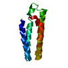
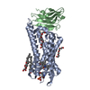
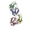
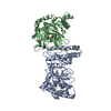
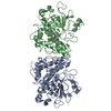
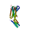
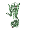
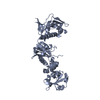
| Title | Crystal structure of the human formyl peptide receptor 2 in complex with WKYMVm | |||||||||||||||
 Components Components |
| |||||||||||||||
 Keywords Keywords | MEMBRANE PROTEIN / Formyl peptide receptor / G protein-coupled receptor / Complex / Peptide agonist | |||||||||||||||
| Function / homology |  Function and homology information Function and homology informationN-formyl peptide receptor activity / complement receptor activity / dentinogenesis / immune response-regulating cell surface receptor signaling pathway / scavenger receptor binding / RAGE receptor binding / complement receptor mediated signaling pathway / positive regulation of monocyte chemotaxis / positive regulation of innate immune response / Formyl peptide receptors bind formyl peptides and many other ligands ...N-formyl peptide receptor activity / complement receptor activity / dentinogenesis / immune response-regulating cell surface receptor signaling pathway / scavenger receptor binding / RAGE receptor binding / complement receptor mediated signaling pathway / positive regulation of monocyte chemotaxis / positive regulation of innate immune response / Formyl peptide receptors bind formyl peptides and many other ligands / cargo receptor activity / positive chemotaxis / tertiary granule membrane / ficolin-1-rich granule membrane / specific granule membrane / positive regulation of superoxide anion generation / positive regulation of phagocytosis / astrocyte activation / receptor-mediated endocytosis / calcium-mediated signaling / microglial cell activation / electron transport chain / negative regulation of inflammatory response / G protein-coupled receptor activity / cellular response to amyloid-beta / chemotaxis / adenylate cyclase-inhibiting G protein-coupled receptor signaling pathway / amyloid-beta binding / positive regulation of cytosolic calcium ion concentration / signaling receptor activity / G alpha (i) signalling events / phospholipase C-activating G protein-coupled receptor signaling pathway / G alpha (q) signalling events / negative regulation of neuron apoptotic process / periplasmic space / positive regulation of ERK1 and ERK2 cascade / electron transfer activity / cell surface receptor signaling pathway / positive regulation of phosphatidylinositol 3-kinase/protein kinase B signal transduction / cell adhesion / defense response to bacterium / iron ion binding / G protein-coupled receptor signaling pathway / inflammatory response / heme binding / Neutrophil degranulation / membrane / plasma membrane / cytoplasm Similarity search - Function | |||||||||||||||
| Biological species |   Homo sapiens (human) Homo sapiens (human)synthetic construct (others) | |||||||||||||||
| Method |  X-RAY DIFFRACTION / SYNCHROTRON / MOLECULAR REPLACEMENT / Resolution: 2.8 Å X-RAY DIFFRACTION / SYNCHROTRON / MOLECULAR REPLACEMENT / Resolution: 2.8 Å | |||||||||||||||
 Authors Authors | Chen, T. / Zong, X. / Zhang, H. / Wang, M. / Zhao, Q. / Wu, B. | |||||||||||||||
| Funding support |  China, 4items China, 4items
| |||||||||||||||
 Citation Citation | Journal: Nat Commun / Year: 2020 Title: Structural basis of ligand binding modes at the human formyl peptide receptor 2. Authors: Chen, T. / Xiong, M. / Zong, X. / Ge, Y. / Zhang, H. / Wang, M. / Won Han, G. / Yi, C. / Ma, L. / Ye, R.D. / Xu, Y. / Zhao, Q. / Wu, B. | |||||||||||||||
| History |
|
- Structure visualization
| Structure viewer | Molecule: MolmilJmol/JSmol |
|---|
- Downloads & links
-Download
| PDBx/mmCIF format | 6lw5.cif.gz | 100.1 KB | Display | PDBx/mmCIF format |
|---|---|---|---|---|
| PDB format | pdb6lw5.ent.gz | 74.5 KB | Display | PDB format |
| PDBx/mmJSON format | 6lw5.json.gz | Tree view | PDBx/mmJSON format | |
| Others |  Other downloads Other downloads |
-Validation report
| Arichive directory | https://data.pdbj.org/pub/pdb/validation_reports/lw/6lw5ftp://data.pdbj.org/pub/pdb/validation_reports/lw/6lw5 | HTTPS FTP |
|---|
-Related structure data


-Links
 PDBj
PDBj


- Assembly
| Deposited unit | 
| ||||||||
|---|---|---|---|---|---|---|---|---|---|
| 1 |
| ||||||||
| Unit cell |
|
-Components
| #1: Protein | Mass: 47694.016 Da / Num. of mol.: 1 / Mutation: M1007W, H1102I, R1106L, S211L Source method: isolated from a genetically manipulated source Source: (gene. exp.) Homo sapiens (human)Gene: cybC, FPR2, FPRH1, FPRL1, LXA4R Production host: Mammalian expression vector Flag-MCS-pcDNA3.1 (others) References: UniProt: P0ABE7, UniProt: P25090 | ||||
|---|---|---|---|---|---|
| #2: Protein/peptide | Mass: 857.117 Da / Num. of mol.: 1 / Source method: obtained synthetically / Source: (synth.) synthetic construct (others) | ||||
| #3: Chemical |   Mass: 386.654 Da / Num. of mol.: 2 / Source method: obtained synthetically / Formula: C27H46O Mass: 386.654 Da / Num. of mol.: 2 / Source method: obtained synthetically / Formula: C27H46O#4: Water | ChemComp-HOH / |  Mass: 18.015 Da / Num. of mol.: 1 / Source method: isolated from a natural source / Formula: H2O Mass: 18.015 Da / Num. of mol.: 1 / Source method: isolated from a natural source / Formula: H2OHas ligand of interest | Y | |
-Experimental details
-Experiment
| Experiment | Method: X-RAY DIFFRACTION / Number of used crystals: 1 |
|---|
- Sample preparation
| Crystal | Density Matthews: 3.26 Å3/Da / Density % sol: 62.22 % |
|---|---|
| Crystal grow | Temperature: 293 K / Method: lipidic cubic phase Details: 0.1 M Tris, pH 7.5, 35% PEG500 DME, 3% PPG400, 100 mM CH3COOLi |
-Data collection
| Diffraction | Mean temperature: 100 K / Serial crystal experiment: N |
|---|---|
| Diffraction source | Source: SYNCHROTRON / Site: SPring-8  / Beamline: BL41XU / Wavelength: 1 Å / Beamline: BL41XU / Wavelength: 1 Å |
| Detector | Type: DECTRIS EIGER X 16M / Detector: PIXEL / Date: Feb 14, 2018 |
| Radiation | Protocol: SINGLE WAVELENGTH / Monochromatic (M) / Laue (L): M / Scattering type: x-ray |
| Radiation wavelength | Wavelength: 1 Å / Relative weight: 1 |
| Reflection | Resolution: 2.8→30 Å / Num. obs: 15644 / % possible obs: 96.5 % / Redundancy: 7.9 % / CC1/2: 0.99 / Net I/σ(I): 6.8 |
| Reflection shell | Resolution: 2.8→2.85 Å / Num. unique obs: 767 / CC1/2: 0.82 |
- Processing
| Software |
| ||||||||||||||||
|---|---|---|---|---|---|---|---|---|---|---|---|---|---|---|---|---|---|
| Refinement | Method to determine structure: MOLECULAR REPLACEMENT Starting model: 5C1M, 1M6T Resolution: 2.8→30 Å / Cross valid method: FREE R-VALUE
| ||||||||||||||||
| Refinement step | Cycle: LAST / Resolution: 2.8→30 Å
| ||||||||||||||||
| LS refinement shell | Resolution: 2.8→2.97 Å
|
