- PDB-6lph: the Sufu-Fu complex crystal structure -
+
データを開く
IDまたはキーワード:
読み込み中...
-
基本情報
登録情報
データベース: PDB / ID: 6lph
タイトル
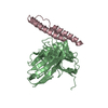
the Sufu-Fu complex crystal structure
要素
Serine/threonine-protein kinase fused
Suppressor of fused homolog
キーワード
PROTEIN BINDING / kinases / phosphorylation
機能・相同性
機能・相同性情報
Hedgehog signaling complex / Assembly of the CI containing complexes / negative regulation of nucleocytoplasmic transport / Activation of CI / Activation of SMO / Hedgehog 'off' state / Phosphorylation of CI / germarium-derived egg chamber formation / Phosphorylation of SMO / Assembly of the 'signalling complexes' ...Hedgehog signaling complex / Assembly of the CI containing complexes / negative regulation of nucleocytoplasmic transport / Activation of CI / Activation of SMO / Hedgehog 'off' state / Phosphorylation of CI / germarium-derived egg chamber formation / Phosphorylation of SMO / Assembly of the 'signalling complexes' / Ubiquitination and proteolysis of phosphorylated CI / Degradation of GLI1 by the proteasome / GLI3 is processed to GLI3R by the proteasome / wing disc pattern formation / Hedgehog 'on' state / positive regulation of nucleocytoplasmic transport / segment polarity determination / smoothened binding / intraciliary transport / nuclear localization sequence binding / smoothened signaling pathway / molecular sequestering activity / negative regulation of BMP signaling pathway / positive regulation of protein ubiquitination / negative regulation of smoothened signaling pathway / DNA-binding transcription factor binding / RNA polymerase II-specific DNA-binding transcription factor binding / non-specific serine/threonine protein kinase / cilium / protein serine kinase activity / protein serine/threonine kinase activity / protein homodimerization activity / protein-containing complex / ATP binding / nucleus / cytoplasm / cytosol 類似検索 - 分子機能
Suppressor of fused / Suppressor of fused, eukaryotic / Suppressor of fused C-terminal / Suppressor of fused, N-terminal / Suppressor of fused, C-terminal domain superfamily / Suppressor of Fused Gli/Ci N terminal binding domain / Suppressor of fused-like domain / Suppressor of fused protein (SUFU) / Serine/threonine-protein kinase, active site / Serine/Threonine protein kinases active-site signature. ...Suppressor of fused / Suppressor of fused, eukaryotic / Suppressor of fused C-terminal / Suppressor of fused, N-terminal / Suppressor of fused, C-terminal domain superfamily / Suppressor of Fused Gli/Ci N terminal binding domain / Suppressor of fused-like domain / Suppressor of fused protein (SUFU) / Serine/threonine-protein kinase, active site / Serine/Threonine protein kinases active-site signature. / Protein kinase domain / Serine/Threonine protein kinases, catalytic domain / Protein kinase, ATP binding site / Protein kinases ATP-binding region signature. / Protein kinase domain profile. / Protein kinase domain / Protein kinase-like domain superfamily 類似検索 - ドメイン・相同性
解像度: 1.91→55.67 Å / Cor.coef. Fo:Fc: 0.963 / Cor.coef. Fo:Fc free: 0.927 / SU B: 7.234 / SU ML: 0.096 / 交差検証法: FREE R-VALUE / σ(F): 0 / ESU R Free: 0.148 詳細: HYDROGENS HAVE BEEN USED IF PRESENT IN THE INPUT U VALUES : WITH TLS ADDED
 ムービー
ムービー コントローラー


 データを開く
 基本情報
基本情報 要素
要素 キーワード
キーワード 機能・相同性情報
 X線回折 /
  データ登録者
 引用
引用 構造の表示
構造の表示 ダウンロードとリンク
 その他のダウンロード


 PDBj
PDBj 集合体
集合体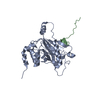

 分子量: 18.015 Da / 分子数: 145 / 由来タイプ: 天然 / 式: H2O
 試料調製
試料調製 / ビームライン: BL17U / 波長: 0.91878 Å
 解析
解析