 Movie
Movie Controller


+ Open data

- Basic information
| Entry | Database: PDB / ID: 6loq | ||||||
|---|---|---|---|---|---|---|---|
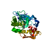
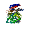
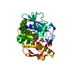
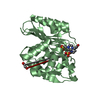
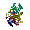
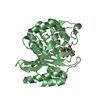
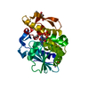
| Title | crystal structure of alpha-momorcharin in complex with cAMP | ||||||
 Components Components | Ribosome-inactivating protein momordin I | ||||||
 Keywords Keywords | PLANT PROTEIN / alpha-momorcharin / Alpha-MMC / ribosome-inactivating protein / rRNA N-glycosidase / cAMP | ||||||
| Function / homology |  Function and homology information Function and homology informationrRNA N-glycosylase / rRNA N-glycosylase activity / defense response / toxin activity / negative regulation of translation Similarity search - Function | ||||||
| Biological species |  Momordica charantia (bitter melon) Momordica charantia (bitter melon) | ||||||
| Method |  X-RAY DIFFRACTION / SYNCHROTRON / MOLECULAR REPLACEMENT / Resolution: 1.331 Å X-RAY DIFFRACTION / SYNCHROTRON / MOLECULAR REPLACEMENT / Resolution: 1.331 Å | ||||||
 Authors Authors | Fan, X. / Jin, T. | ||||||
 Citation Citation | Journal: Int.J.Biol.Macromol. / Year: 2020 Title: Atomic-resolution structures of type I ribosome inactivating protein alpha-momorcharin with different substrate analogs. Authors: Fan, X. / Wang, Y. / Guo, F. / Zhang, Y. / Jin, T. | ||||||
| History |
|
- Structure visualization
| Structure viewer | Molecule: MolmilJmol/JSmol |
|---|
- Downloads & links
-Download
| PDBx/mmCIF format | 6loq.cif.gz | 107.9 KB | Display | PDBx/mmCIF format |
|---|---|---|---|---|
| PDB format | pdb6loq.ent.gz | 77.9 KB | Display | PDB format |
| PDBx/mmJSON format | 6loq.json.gz | Tree view | PDBx/mmJSON format | |
| Others |  Other downloads Other downloads |
-Validation report
| Arichive directory | https://data.pdbj.org/pub/pdb/validation_reports/lo/6loqftp://data.pdbj.org/pub/pdb/validation_reports/lo/6loq | HTTPS FTP |
|---|
-Related structure data
| Related structure data |  6lorC  6lovC 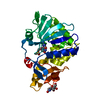 6lowC  6loyC  6lozC  6lp0C  1f8qS S: Starting model for refinement C: citing same article ( |
|---|---|
| Similar structure data |


-Links
 PDBj
PDBj


- Assembly
| Deposited unit | 
| ||||||||
|---|---|---|---|---|---|---|---|---|---|
| 1 |
| ||||||||
| Unit cell |
|
-Components
| #1: Protein | Mass: 31562.961 Da / Num. of mol.: 1 / Source method: isolated from a natural source / Details: cAMP / Source: (natural) Momordica charantia (bitter melon) / References: UniProt: P16094, rRNA N-glycosylase |
|---|---|
| #2: Chemical | ChemComp-CMP /   Mass: 329.206 Da / Num. of mol.: 1 / Source method: obtained synthetically / Formula: C10H12N5O6P / Feature type: SUBJECT OF INVESTIGATION Mass: 329.206 Da / Num. of mol.: 1 / Source method: obtained synthetically / Formula: C10H12N5O6P / Feature type: SUBJECT OF INVESTIGATION |
| #3: Water | ChemComp-HOH /  Mass: 18.015 Da / Num. of mol.: 196 / Source method: isolated from a natural source / Formula: H2O Mass: 18.015 Da / Num. of mol.: 196 / Source method: isolated from a natural source / Formula: H2O |
| Has ligand of interest | Y |
-Experimental details
-Experiment
| Experiment | Method: X-RAY DIFFRACTION / Number of used crystals: 1 |
|---|
- Sample preparation
| Crystal | Density Matthews: 2.06 Å3/Da / Density % sol: 40.26 % |
|---|---|
| Crystal grow | Temperature: 298 K / Method: vapor diffusion, hanging drop / pH: 6.5 Details: 0.2 M Magnesium Acetate, 20% PEG 8000, 0.1 M Sodium cacodylate, pH 6.5 |
-Data collection
| Diffraction | Mean temperature: 100 K / Serial crystal experiment: N |
|---|---|
| Diffraction source | Source: SYNCHROTRON / Site: APS  / Beamline: 22-ID / Wavelength: 1 Å / Beamline: 22-ID / Wavelength: 1 Å |
| Detector | Type: MARMOSAIC 300 mm CCD / Detector: CCD / Date: Dec 6, 2007 |
| Radiation | Protocol: SINGLE WAVELENGTH / Monochromatic (M) / Laue (L): M / Scattering type: x-ray |
| Radiation wavelength | Wavelength: 1 Å / Relative weight: 1 |
| Reflection | Resolution: 1.33→50 Å / Num. obs: 55352 / % possible obs: 96 % / Redundancy: 5 % / CC1/2: 0.992 / Rmerge(I) obs: 0.08 / Rpim(I) all: 0.044 / Rrim(I) all: 0.092 / Net I/σ(I): 14.2 |
| Reflection shell | Resolution: 1.33→1.38 Å / Rmerge(I) obs: 0.943 / Mean I/σ(I) obs: 1.6 / Num. unique obs: 5700 / CC1/2: 0.683 / Rpim(I) all: 0.46 |
- Processing
| Software |
| ||||||||||||||||||||||||||||||||||||||||||||||||||||||||||||||||||||||||||||||||||||||||||||||||||||||||||||||||||||||||||||||||||||||||||||||||||||||
|---|---|---|---|---|---|---|---|---|---|---|---|---|---|---|---|---|---|---|---|---|---|---|---|---|---|---|---|---|---|---|---|---|---|---|---|---|---|---|---|---|---|---|---|---|---|---|---|---|---|---|---|---|---|---|---|---|---|---|---|---|---|---|---|---|---|---|---|---|---|---|---|---|---|---|---|---|---|---|---|---|---|---|---|---|---|---|---|---|---|---|---|---|---|---|---|---|---|---|---|---|---|---|---|---|---|---|---|---|---|---|---|---|---|---|---|---|---|---|---|---|---|---|---|---|---|---|---|---|---|---|---|---|---|---|---|---|---|---|---|---|---|---|---|---|---|---|---|---|---|---|---|
| Refinement | Method to determine structure: MOLECULAR REPLACEMENT Starting model: 1F8Q Resolution: 1.331→24.604 Å / Cor.coef. Fo:Fc: 0.95 / Cor.coef. Fo:Fc free: 0.857 / WRfactor Rfree: 0.279 / WRfactor Rwork: 0.236 / SU B: 2.801 / SU ML: 0.053 / Average fsc free: 0.8302 / Average fsc work: 0.8321 / Cross valid method: FREE R-VALUE / ESU R: 0.066 / ESU R Free: 0.071 Details: Hydrogens have been added in their riding positions
| ||||||||||||||||||||||||||||||||||||||||||||||||||||||||||||||||||||||||||||||||||||||||||||||||||||||||||||||||||||||||||||||||||||||||||||||||||||||
| Solvent computation | Ion probe radii: 0.8 Å / Shrinkage radii: 0.8 Å / VDW probe radii: 1.2 Å / Solvent model: MASK BULK SOLVENT | ||||||||||||||||||||||||||||||||||||||||||||||||||||||||||||||||||||||||||||||||||||||||||||||||||||||||||||||||||||||||||||||||||||||||||||||||||||||
| Displacement parameters | Biso mean: 25.452 Å2
| ||||||||||||||||||||||||||||||||||||||||||||||||||||||||||||||||||||||||||||||||||||||||||||||||||||||||||||||||||||||||||||||||||||||||||||||||||||||
| Refinement step | Cycle: LAST / Resolution: 1.331→24.604 Å
| ||||||||||||||||||||||||||||||||||||||||||||||||||||||||||||||||||||||||||||||||||||||||||||||||||||||||||||||||||||||||||||||||||||||||||||||||||||||
| Refine LS restraints |
| ||||||||||||||||||||||||||||||||||||||||||||||||||||||||||||||||||||||||||||||||||||||||||||||||||||||||||||||||||||||||||||||||||||||||||||||||||||||
| LS refinement shell |
| ||||||||||||||||||||||||||||||||||||||||||||||||||||||||||||||||||||||||||||||||||||||||||||||||||||||||||||||||||||||||||||||||||||||||||||||||||||||
| Refinement TLS params. | Method: refined / Origin x: -20.4672 Å / Origin y: -12.6721 Å / Origin z: 0.0318 Å
| ||||||||||||||||||||||||||||||||||||||||||||||||||||||||||||||||||||||||||||||||||||||||||||||||||||||||||||||||||||||||||||||||||||||||||||||||||||||
| Refinement TLS group | Selection: ALL |
