 Movie
Movie Controller

[English] 日本語
 Yorodumi
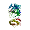
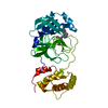
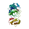
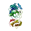
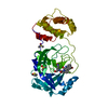
- PDB-6lny: The co-crystal structure of Severe Acute Respiratory Syndrome Cor... -
+ Open data

- Basic information
| Entry | Database: PDB / ID: 6lny | ||||||
|---|---|---|---|---|---|---|---|
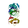
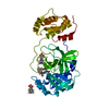
| Title | The co-crystal structure of Severe Acute Respiratory Syndrome Coronavirus 3C-Like Protease with aldehyde M15 | ||||||
 Components Components | Replicase polyprotein 1a | ||||||
 Keywords Keywords | HYDROLASE / Severe Acute Respiratory Syndrome Coronavirus / 3C Like Protease | ||||||
| Function / homology |  Function and homology information Function and homology informationAssembly of the SARS-CoV-1 Replication-Transcription Complex (RTC) / Maturation of replicase proteins / Transcription of SARS-CoV-1 sgRNAs / Translation of Replicase and Assembly of the Replication Transcription Complex / K48-linked deubiquitinase activity / Replication of the SARS-CoV-1 genome / K63-linked deubiquitinase activity / host cell endoplasmic reticulum / SARS-CoV-1 modulates host translation machinery / viral genome replication ...Assembly of the SARS-CoV-1 Replication-Transcription Complex (RTC) / Maturation of replicase proteins / Transcription of SARS-CoV-1 sgRNAs / Translation of Replicase and Assembly of the Replication Transcription Complex / K48-linked deubiquitinase activity / Replication of the SARS-CoV-1 genome / K63-linked deubiquitinase activity / host cell endoplasmic reticulum / SARS-CoV-1 modulates host translation machinery / viral genome replication / methyltransferase activity / SARS-CoV-1 activates/modulates innate immune responses / endonuclease activity / methylation / double membrane vesicle viral factory outer membrane / SARS coronavirus main proteinase / host cell endosome / symbiont-mediated degradation of host mRNA / mRNA guanylyltransferase / symbiont-mediated suppression of host ISG15-protein conjugation / G-quadruplex RNA binding / mRNA guanylyltransferase activity / symbiont-mediated suppression of host cytoplasmic pattern recognition receptor signaling pathway via inhibition of IRF3 activity / omega peptidase activity / symbiont-mediated perturbation of host ubiquitin-like protein modification / host cell Golgi apparatus / ubiquitinyl hydrolase 1 / cysteine-type deubiquitinase activity / Hydrolases; Acting on peptide bonds (peptidases); Cysteine endopeptidases / lyase activity / single-stranded RNA binding / viral protein processing / host cell perinuclear region of cytoplasm / symbiont-mediated suppression of host type I interferon-mediated signaling pathway / symbiont-mediated suppression of host gene expression / viral translational frameshifting / symbiont-mediated activation of host autophagy / cysteine-type endopeptidase activity / RNA-directed RNA polymerase activity / proteolysis / zinc ion binding / identical protein binding Similarity search - Function | ||||||
| Biological species |   Human SARS coronavirus Human SARS coronavirus | ||||||
| Method |  X-RAY DIFFRACTION / MOLECULAR REPLACEMENT / Resolution: 2.245 Å X-RAY DIFFRACTION / MOLECULAR REPLACEMENT / Resolution: 2.245 Å | ||||||
 Authors Authors | Wang, H. / Shang, L.Q. | ||||||
| Funding support |  China, 1items China, 1items
| ||||||
 Citation Citation | Journal: Acs Catalysis / Year: 2020 Title: Comprehensive Insights into the Catalytic Mechanism of Middle East Respiratory Syndrome 3C-Like Protease and Severe Acute Respiratory Syndrome 3C-Like Protease. Authors: Wang, H. / He, S. / Deng, W. / Zhang, Y. / Li, G. / Sun, J. / Zhao, W. / Guo, Y. / Yin, Z. / Li, D. / Shang, L. | ||||||
| History |
|
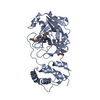
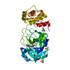
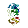
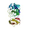
- Structure visualization
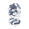
| Structure viewer | Molecule: MolmilJmol/JSmol |
|---|
- Downloads & links
-Download
| PDBx/mmCIF format | 6lny.cif.gz | 77.1 KB | Display | PDBx/mmCIF format |
|---|---|---|---|---|
| PDB format | pdb6lny.ent.gz | 55.4 KB | Display | PDB format |
| PDBx/mmJSON format | 6lny.json.gz | Tree view | PDBx/mmJSON format | |
| Others |  Other downloads Other downloads |
-Validation report
| Arichive directory | https://data.pdbj.org/pub/pdb/validation_reports/ln/6lnyftp://data.pdbj.org/pub/pdb/validation_reports/ln/6lny | HTTPS FTP |
|---|
-Related structure data
| Related structure data |  6lnqC 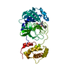 6lo0C 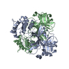 1uj1S S: Starting model for refinement C: citing same article ( |
|---|---|
| Similar structure data |


-Links
 PDBj
PDBj


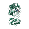
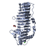
- Assembly
| Deposited unit | 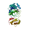
| |||||||||
|---|---|---|---|---|---|---|---|---|---|---|
| 1 |
| |||||||||
| Unit cell |
| |||||||||
| Components on special symmetry positions |
|
-Components
| #1: Protein | Mass: 33876.637 Da / Num. of mol.: 1 Source method: isolated from a genetically manipulated source Source: (gene. exp.) Human SARS coronavirus / Gene: 1a / Production host:  References: UniProt: P0C6U8, ubiquitinyl hydrolase 1, SARS coronavirus main proteinase, Hydrolases; Acting on peptide bonds (peptidases); Cysteine endopeptidases |
|---|---|
| #2: Chemical | ChemComp-EOC / (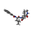  Mass: 413.510 Da / Num. of mol.: 1 / Source method: obtained synthetically / Formula: C23H31N3O4 / Feature type: SUBJECT OF INVESTIGATION Mass: 413.510 Da / Num. of mol.: 1 / Source method: obtained synthetically / Formula: C23H31N3O4 / Feature type: SUBJECT OF INVESTIGATION |
| #3: Water | ChemComp-HOH /  Mass: 18.015 Da / Num. of mol.: 109 / Source method: isolated from a natural source / Formula: H2O Mass: 18.015 Da / Num. of mol.: 109 / Source method: isolated from a natural source / Formula: H2O |
| Has ligand of interest | Y |
-Experimental details
-Experiment
| Experiment | Method: X-RAY DIFFRACTION / Number of used crystals: 1 |
|---|
- Sample preparation
| Crystal | Density Matthews: 3.36 Å3/Da / Density % sol: 63.36 % |
|---|---|
| Crystal grow | Temperature: 289 K / Method: vapor diffusion, hanging drop / pH: 6 / Details: PEG 8000, 100mM MES buffer |
-Data collection
| Diffraction | Mean temperature: 100 K / Serial crystal experiment: N |
|---|---|
| Diffraction source | Source: ROTATING ANODE / Type: RIGAKU RU200 / Wavelength: 1.5418 Å |
| Detector | Type: RIGAKU RAXIS IV++ / Detector: IMAGE PLATE / Date: Jan 2, 2019 |
| Radiation | Protocol: SINGLE WAVELENGTH / Monochromatic (M) / Laue (L): M / Scattering type: x-ray |
| Radiation wavelength | Wavelength: 1.5418 Å / Relative weight: 1 |
| Reflection | Resolution: 2.245→50 Å / Num. obs: 21495 / % possible obs: 98.7 % / Redundancy: 6.5 % / Rmerge(I) obs: 0.09 / Net I/σ(I): 21.68 |
| Reflection shell | Resolution: 2.245→2.28 Å / Rmerge(I) obs: 0.318 / Num. unique obs: 930 |
- Processing
| Software |
| ||||||||||||||||||||||||||||||||||||||||||||||||||||||||||||||||||||||||||||||||||||||||||
|---|---|---|---|---|---|---|---|---|---|---|---|---|---|---|---|---|---|---|---|---|---|---|---|---|---|---|---|---|---|---|---|---|---|---|---|---|---|---|---|---|---|---|---|---|---|---|---|---|---|---|---|---|---|---|---|---|---|---|---|---|---|---|---|---|---|---|---|---|---|---|---|---|---|---|---|---|---|---|---|---|---|---|---|---|---|---|---|---|---|---|---|
| Refinement | Method to determine structure: MOLECULAR REPLACEMENT Starting model: 1UJ1 Resolution: 2.245→40.335 Å / SU ML: 0.31 / Cross valid method: THROUGHOUT / σ(F): 1.34 / Phase error: 29.37
| ||||||||||||||||||||||||||||||||||||||||||||||||||||||||||||||||||||||||||||||||||||||||||
| Solvent computation | Shrinkage radii: 0.9 Å / VDW probe radii: 1.11 Å | ||||||||||||||||||||||||||||||||||||||||||||||||||||||||||||||||||||||||||||||||||||||||||
| Displacement parameters | Biso max: 83.77 Å2 / Biso mean: 32.9825 Å2 / Biso min: 16.81 Å2 | ||||||||||||||||||||||||||||||||||||||||||||||||||||||||||||||||||||||||||||||||||||||||||
| Refinement step | Cycle: final / Resolution: 2.245→40.335 Å
| ||||||||||||||||||||||||||||||||||||||||||||||||||||||||||||||||||||||||||||||||||||||||||
| LS refinement shell | Refine-ID: X-RAY DIFFRACTION / Rfactor Rfree error: 0
|
