 Movie
Movie Controller

[English] 日本語
 Yorodumi
- PDB-6n2b: The Crystal Structure of Caldicellulosiruptor kristjanssonii Tapi... -
+ Open data

- Basic information
| Entry | Database: PDB / ID: 6n2b | ||||||
|---|---|---|---|---|---|---|---|
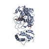
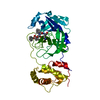
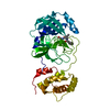
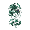
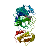
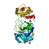
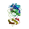
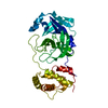
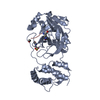
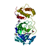
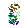
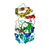
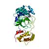
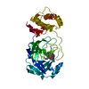
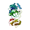
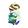
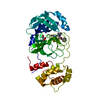
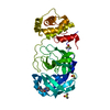
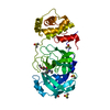
| Title | The Crystal Structure of Caldicellulosiruptor kristjanssonii Tapirin C-terminal domain | ||||||
 Components Components | Tapirin | ||||||
 Keywords Keywords | CELL ADHESION / BETA-HELIX / cellulose binding | ||||||
| Function / homology | membrane / metal ion binding / Type 4 fimbrial biogenesis protein PilX N-terminal domain-containing protein Function and homology information Function and homology information | ||||||
| Biological species | Caldicellulosiruptor kristjanssonii | ||||||
| Method |  X-RAY DIFFRACTION / MOLECULAR REPLACEMENT / Resolution: 2.65 Å X-RAY DIFFRACTION / MOLECULAR REPLACEMENT / Resolution: 2.65 Å | ||||||
 Authors Authors | Alahuhta, P.M. / Lunin, V.V. | ||||||
| Funding support |  United States, 1items United States, 1items
| ||||||
 Citation Citation | Journal: Appl. Environ. Microbiol. / Year: 2019 Title: Comparative Biochemical and Structural Analysis of Novel Cellulose Binding Proteins (Tapirins) from Extremely ThermophilicCaldicellulosiruptorSpecies. Authors: Lee, L.L. / Hart, W.S. / Lunin, V.V. / Alahuhta, M. / Bomble, Y.J. / Himmel, M.E. / Blumer-Schuette, S.E. / Adams, M.W.W. / Kelly, R.M. | ||||||
| History |
|
- Structure visualization
| Structure viewer | Molecule: MolmilJmol/JSmol |
|---|
- Downloads & links
-Download
| PDBx/mmCIF format | 6n2b.cif.gz | 170.4 KB | Display | PDBx/mmCIF format |
|---|---|---|---|---|
| PDB format | pdb6n2b.ent.gz | 128.1 KB | Display | PDB format |
| PDBx/mmJSON format | 6n2b.json.gz | Tree view | PDBx/mmJSON format | |
| Others |  Other downloads Other downloads |
-Validation report
| Arichive directory | https://data.pdbj.org/pub/pdb/validation_reports/n2/6n2bftp://data.pdbj.org/pub/pdb/validation_reports/n2/6n2b | HTTPS FTP |
|---|
-Related structure data
| Related structure data | 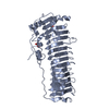 6n2cC 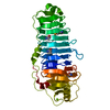 4wa0S S: Starting model for refinement C: citing same article ( |
|---|---|
| Similar structure data |
-Links
 PDBj
PDBj- Assembly
| Deposited unit | 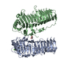
| ||||||||
|---|---|---|---|---|---|---|---|---|---|
| 1 | 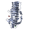
| ||||||||
| 2 | 
| ||||||||
| Unit cell |
|
-Components
| #1: Protein | Mass: 62925.223 Da / Num. of mol.: 2 / Fragment: C-terminal domain residues 59-634 Source method: isolated from a genetically manipulated source Source: (gene. exp.)  Caldicellulosiruptor kristjanssonii (strain ATCC 700853 / DSM 12137 / I77R1B) (bacteria) Caldicellulosiruptor kristjanssonii (strain ATCC 700853 / DSM 12137 / I77R1B) (bacteria)Strain: ATCC 700853 / DSM 12137 / I77R1B / Gene: Calkr_0826 / Production host: #2: Chemical |   Mass: 40.078 Da / Num. of mol.: 2 / Source method: obtained synthetically / Formula: Ca Mass: 40.078 Da / Num. of mol.: 2 / Source method: obtained synthetically / Formula: Ca#3: Chemical | ChemComp-GOL / |   Mass: 92.094 Da / Num. of mol.: 1 / Source method: obtained synthetically / Formula: C3H8O3 Mass: 92.094 Da / Num. of mol.: 1 / Source method: obtained synthetically / Formula: C3H8O3#4: Water | ChemComp-HOH / |  Mass: 18.015 Da / Num. of mol.: 629 / Source method: isolated from a natural source / Formula: H2O Mass: 18.015 Da / Num. of mol.: 629 / Source method: isolated from a natural source / Formula: H2O |
|---|
-Experimental details
-Experiment
| Experiment | Method: X-RAY DIFFRACTION / Number of used crystals: 1 |
|---|
- Sample preparation
| Crystal | Density Matthews: 2.57 Å3/Da / Density % sol: 52.12 % |
|---|---|
| Crystal grow | Temperature: 293 K / Method: vapor diffusion, sitting drop Details: 0.1 M citric acid pH 3.0 to 4.0 and 10% to 15% w/v polyethylene glycol 3350 |
-Data collection
| Diffraction | Mean temperature: 100 K / Serial crystal experiment: N |
|---|---|
| Diffraction source | Source: ROTATING ANODE / Type: BRUKER AXS MICROSTAR / Wavelength: 1.5418 Å |
| Detector | Type: Bruker Platinum 135 / Detector: CCD / Date: Aug 21, 2017 / Details: HELIOS MIRRORS |
| Radiation | Protocol: SINGLE WAVELENGTH / Monochromatic (M) / Laue (L): M / Scattering type: x-ray |
| Radiation wavelength | Wavelength: 1.5418 Å / Relative weight: 1 |
| Reflection | Resolution: 2.65→55 Å / Num. obs: 38585 / % possible obs: 100 % / Redundancy: 5.23 % / Rsym value: 0.2419 / Net I/σ(I): 4.89 |
| Reflection shell | Resolution: 2.65→2.75 Å / Redundancy: 4.26 % / Mean I/σ(I) obs: 1.03 / Num. unique obs: 3969 / Rsym value: 0.6461 / % possible all: 100 |
- Processing
| Software |
| |||||||||||||||||||||||||||||||||||||||||||||||||||||||||||||||||||||||||||||||||||||||||||||||||||||||||
|---|---|---|---|---|---|---|---|---|---|---|---|---|---|---|---|---|---|---|---|---|---|---|---|---|---|---|---|---|---|---|---|---|---|---|---|---|---|---|---|---|---|---|---|---|---|---|---|---|---|---|---|---|---|---|---|---|---|---|---|---|---|---|---|---|---|---|---|---|---|---|---|---|---|---|---|---|---|---|---|---|---|---|---|---|---|---|---|---|---|---|---|---|---|---|---|---|---|---|---|---|---|---|---|---|---|---|
| Refinement | Method to determine structure: MOLECULAR REPLACEMENT Starting model: 4WA0 Resolution: 2.65→54.425 Å / SU ML: 0.39 / Cross valid method: FREE R-VALUE / σ(F): 1.34 / Phase error: 24.66
| |||||||||||||||||||||||||||||||||||||||||||||||||||||||||||||||||||||||||||||||||||||||||||||||||||||||||
| Solvent computation | Shrinkage radii: 0.9 Å / VDW probe radii: 1.11 Å | |||||||||||||||||||||||||||||||||||||||||||||||||||||||||||||||||||||||||||||||||||||||||||||||||||||||||
| Refinement step | Cycle: LAST / Resolution: 2.65→54.425 Å
| |||||||||||||||||||||||||||||||||||||||||||||||||||||||||||||||||||||||||||||||||||||||||||||||||||||||||
| Refine LS restraints |
| |||||||||||||||||||||||||||||||||||||||||||||||||||||||||||||||||||||||||||||||||||||||||||||||||||||||||
| LS refinement shell |
|
