 Movie
Movie Controller

[English] 日本語
 Yorodumi
- PDB-6lni: Cryo-EM structure of amyloid fibril formed by full-length human p... -
+ Open data

- Basic information
| Entry | Database: PDB / ID: 6lni | ||||||
|---|---|---|---|---|---|---|---|
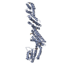
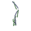
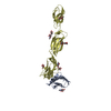
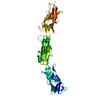
| Title | Cryo-EM structure of amyloid fibril formed by full-length human prion protein | ||||||
 Components Components | Major prion protein | ||||||
 Keywords Keywords | PROTEIN FIBRIL / Amyloid fibril | ||||||
| Function / homology |  Function and homology information Function and homology informationnegative regulation of amyloid precursor protein catabolic process / regulation of glutamate receptor signaling pathway / lamin binding / aspartic-type endopeptidase inhibitor activity / regulation of calcium ion import across plasma membrane / glycosaminoglycan binding / NCAM1 interactions / positive regulation of glutamate receptor signaling pathway / negative regulation of interleukin-17 production / cupric ion binding ...negative regulation of amyloid precursor protein catabolic process / regulation of glutamate receptor signaling pathway / lamin binding / aspartic-type endopeptidase inhibitor activity / regulation of calcium ion import across plasma membrane / glycosaminoglycan binding / NCAM1 interactions / positive regulation of glutamate receptor signaling pathway / negative regulation of interleukin-17 production / cupric ion binding / ATP-dependent protein binding / negative regulation of protein processing / regulation of potassium ion transmembrane transport / type 5 metabotropic glutamate receptor binding / negative regulation of dendritic spine maintenance / dendritic spine maintenance / extrinsic component of membrane / negative regulation of calcineurin-NFAT signaling cascade / negative regulation of interleukin-2 production / Insertion of tail-anchored proteins into the endoplasmic reticulum membrane / negative regulation of activated T cell proliferation / negative regulation of amyloid-beta formation / response to amyloid-beta / negative regulation of type II interferon production / cuprous ion binding / negative regulation of long-term synaptic potentiation / intracellular copper ion homeostasis / negative regulation of T cell receptor signaling pathway / positive regulation of protein targeting to membrane / response to cadmium ion / long-term memory / neuron projection maintenance / inclusion body / positive regulation of calcium-mediated signaling / molecular function activator activity / cellular response to copper ion / positive regulation of protein localization to plasma membrane / molecular condensate scaffold activity / tubulin binding / protein homooligomerization / protein destabilization / cellular response to xenobiotic stimulus / cellular response to amyloid-beta / terminal bouton / positive regulation of neuron apoptotic process / amyloid-beta binding / protein-folding chaperone binding / signaling receptor activity / protease binding / response to oxidative stress / nuclear membrane / microtubule binding / molecular adaptor activity / transmembrane transporter binding / learning or memory / regulation of cell cycle / postsynaptic density / intracellular signal transduction / postsynapse / membrane raft / copper ion binding / external side of plasma membrane / dendrite / negative regulation of apoptotic process / protein-containing complex binding / cell surface / negative regulation of transcription by RNA polymerase II / Golgi apparatus / endoplasmic reticulum / extracellular exosome / identical protein binding / plasma membrane / cytoplasm / cytosol Similarity search - Function | ||||||
| Biological species |  Homo sapiens (human) Homo sapiens (human) | ||||||
| Method | ELECTRON MICROSCOPY / helical reconstruction / cryo EM / Resolution: 2.702 Å | ||||||
 Authors Authors | Wang, L.Q. / Zhao, K. / Yuan, H.Y. / Wang, Q. / Guan, Z.Y. / Tao, J. / Li, X.N. / Hao, M.M. / Chen, J. / Zhang, D.L. ...Wang, L.Q. / Zhao, K. / Yuan, H.Y. / Wang, Q. / Guan, Z.Y. / Tao, J. / Li, X.N. / Hao, M.M. / Chen, J. / Zhang, D.L. / Zhu, H.L. / Yin, P. / Liu, C. / Liang, Y. | ||||||
 Citation Citation | Journal: Nat Struct Mol Biol / Year: 2020 Title: Cryo-EM structure of an amyloid fibril formed by full-length human prion protein. Authors: Li-Qiang Wang / Kun Zhao / Han-Ye Yuan / Qiang Wang / Zeyuan Guan / Jing Tao / Xiang-Ning Li / Yunpeng Sun / Chuan-Wei Yi / Jie Chen / Dan Li / Delin Zhang / Ping Yin / Cong Liu / Yi Liang /  Abstract: Prion diseases are caused by the misfolding of prion protein (PrP). Misfolded PrP forms protease-resistant aggregates in vivo (PrP) that are able to template the conversion of the native form of the ...Prion diseases are caused by the misfolding of prion protein (PrP). Misfolded PrP forms protease-resistant aggregates in vivo (PrP) that are able to template the conversion of the native form of the protein (PrP), a property shared by in vitro-produced PrP fibrils. Here we produced amyloid fibrils in vitro from recombinant, full-length human PrP (residues 23-231) and determined their structure using cryo-EM, building a model for the fibril core comprising residues 170-229. The PrP fibril consists of two protofibrils intertwined in a left-handed helix. Lys194 and Glu196 from opposing subunits form salt bridges, creating a hydrophilic cavity at the interface of the two protofibrils. By comparison with the structure of PrP, we propose that two α-helices in the C-terminal domain of PrP are converted into β-strands stabilized by a disulfide bond in the PrP fibril. Our data suggest that different PrP mutations may play distinct roles in modulating the conformational conversion. | ||||||
| History |
|
- Structure visualization
| Movie |
Movie viewer |
|---|---|
| Structure viewer | Molecule: MolmilJmol/JSmol |
- Downloads & links
-Download
| PDBx/mmCIF format | 6lni.cif.gz | 134.4 KB | Display | PDBx/mmCIF format |
|---|---|---|---|---|
| PDB format | pdb6lni.ent.gz | 96.6 KB | Display | PDB format |
| PDBx/mmJSON format | 6lni.json.gz | Tree view | PDBx/mmJSON format | |
| Others |  Other downloads Other downloads |
-Validation report
| Arichive directory | https://data.pdbj.org/pub/pdb/validation_reports/ln/6lniftp://data.pdbj.org/pub/pdb/validation_reports/ln/6lni | HTTPS FTP |
|---|
-Related structure data
| Related structure data |  0931MC M: map data used to model this data C: citing same article ( |
|---|---|
| Similar structure data |


-Links
 PDBj
PDBj


- Assembly
| Deposited unit | 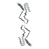
|
|---|---|
| 1 |
|
-Components
| #1: Protein | Mass: 22996.355 Da / Num. of mol.: 10 Source method: isolated from a genetically manipulated source Source: (gene. exp.) Homo sapiens (human) / Gene: PRNP, ALTPRP, PRIP, PRP / Production host:  Has protein modification | Y | Sequence details | Regarding the conflict, the Met residue is a part of the initiation codon of human prion protein ...Regarding the conflict, the Met residue is a part of the initiation codon of human prion protein (PrP) in prokaryotic expression system. (see SEQADV) | |
|---|
-Experimental details
-Experiment
| Experiment | Method: ELECTRON MICROSCOPY |
|---|---|
| EM experiment | Aggregation state: HELICAL ARRAY / 3D reconstruction method: helical reconstruction |
- Sample preparation
| Component | Name: human prion protein amyloid fibril / Type: ORGANELLE OR CELLULAR COMPONENT / Entity ID: all / Source: RECOMBINANT |
|---|---|
| Source (natural) | Organism: Homo sapiens (human) |
| Source (recombinant) | Organism: |
| Buffer solution | pH: 5 |
| Specimen | Embedding applied: NO / Shadowing applied: NO / Staining applied: NO / Vitrification applied: YES |
| Vitrification | Cryogen name: ETHANE |
- Electron microscopy imaging
| Experimental equipment |  Model: Titan Krios / Image courtesy: FEI Company |
|---|---|
| Microscopy | Model: FEI TITAN KRIOS |
| Electron gun | Electron source:  FIELD EMISSION GUN / Accelerating voltage: 300 kV / Illumination mode: FLOOD BEAM FIELD EMISSION GUN / Accelerating voltage: 300 kV / Illumination mode: FLOOD BEAM |
| Electron lens | Mode: BRIGHT FIELD |
| Image recording | Electron dose: 64 e/Å2 / Film or detector model: GATAN K2 QUANTUM (4k x 4k) |
- Processing
| CTF correction | Type: NONE |
|---|---|
| Helical symmerty | Angular rotation/subunit: 179.449 ° / Axial rise/subunit: 2.403 Å / Axial symmetry: C1 |
| 3D reconstruction | Resolution: 2.702 Å / Resolution method: FSC 0.143 CUT-OFF / Num. of particles: 86012 / Symmetry type: HELICAL |
