 Movie
Movie Controller

[English] 日本語
 Yorodumi
- PDB-6li9: Heteromeric amino acid transporter b0,+AT-rBAT complex bound with... -
+ Open data

- Basic information
| Entry | Database: PDB / ID: 6li9 | ||||||||||||||||||||||||
|---|---|---|---|---|---|---|---|---|---|---|---|---|---|---|---|---|---|---|---|---|---|---|---|---|---|
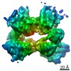
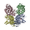
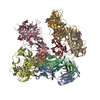
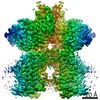
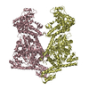
| Title | Heteromeric amino acid transporter b0,+AT-rBAT complex bound with Arginine | ||||||||||||||||||||||||
 Components Components |
| ||||||||||||||||||||||||
 Keywords Keywords | MEMBRANE PROTEIN / transporter | ||||||||||||||||||||||||
| Function / homology |  Function and homology information Function and homology informationbroad specificity neutral L-amino acid:basic L-amino acid antiporter activity / Defective SLC3A1 causes cystinuria (CSNU) / Defective amino acid transport by SLC7A9 causes cystinuria (CSNU) / basic amino acid transport / L-cystine transmembrane transporter activity / basic amino acid transmembrane transporter activity / L-cystine transport / aspartate transmembrane transport / amino acid transmembrane transport / L-glutamate transmembrane transport ...broad specificity neutral L-amino acid:basic L-amino acid antiporter activity / Defective SLC3A1 causes cystinuria (CSNU) / Defective amino acid transport by SLC7A9 causes cystinuria (CSNU) / basic amino acid transport / L-cystine transmembrane transporter activity / basic amino acid transmembrane transporter activity / L-cystine transport / aspartate transmembrane transport / amino acid transmembrane transport / L-glutamate transmembrane transport / neutral amino acid transport / neutral L-amino acid transmembrane transporter activity / Amino acid transport across the plasma membrane / amino acid transmembrane transporter activity / antiporter activity / vacuolar membrane / Basigin interactions / amino acid transport / brush border membrane / peptide antigen binding / protein-containing complex assembly / carbohydrate metabolic process / gene expression / apical plasma membrane / protein heterodimerization activity / protein-containing complex binding / extracellular exosome / membrane / plasma membrane Similarity search - Function | ||||||||||||||||||||||||
| Biological species |  Homo sapiens (human) Homo sapiens (human) | ||||||||||||||||||||||||
| Method | ELECTRON MICROSCOPY / single particle reconstruction / cryo EM / Resolution: 2.3 Å | ||||||||||||||||||||||||
 Authors Authors | Yan, R.H. / Li, Y.N. / Lei, J.L. / Zhou, Q. | ||||||||||||||||||||||||
| Funding support |  China, 7items China, 7items
| ||||||||||||||||||||||||
 Citation Citation | Journal: Sci Adv / Year: 2020 Title: Cryo-EM structure of the human heteromeric amino acid transporter bAT-rBAT. Authors: Renhong Yan / Yaning Li / Yi Shi / Jiayao Zhou / Jianlin Lei / Jing Huang / Qiang Zhou / Abstract: Heteromeric amino acid transporters (HATs) catalyze the transmembrane movement of amino acids, comprising two subunits, a heavy chain and a light chain, linked by a disulfide bridge. The bAT (SLC7A9) ...Heteromeric amino acid transporters (HATs) catalyze the transmembrane movement of amino acids, comprising two subunits, a heavy chain and a light chain, linked by a disulfide bridge. The bAT (SLC7A9) is a representative light chain of HATs, forming heterodimer with rBAT, a heavy chain which mediates the membrane trafficking of bAT. The bAT-rBAT complex is an obligatory exchanger, which mediates the influx of cystine and cationic amino acids and the efflux of neutral amino acids in kidney and small intestine. Here, we report the cryo-EM structure of the human bAT-rBAT complex alone and in complex with arginine substrate at resolution of 2.7 and 2.3 Å, respectively. The overall structure of bAT-rBAT exists as a dimer of heterodimer consistent with the previous study. A ligand molecule is bound to the substrate binding pocket, near which an occluded pocket is identified, to which we found that it is important for substrate transport. | ||||||||||||||||||||||||
| History |
|
- Structure visualization
| Movie |
Movie viewer |
|---|---|
| Structure viewer | Molecule: MolmilJmol/JSmol |
- Downloads & links
-Download
| PDBx/mmCIF format | 6li9.cif.gz | 411 KB | Display | PDBx/mmCIF format |
|---|---|---|---|---|
| PDB format | pdb6li9.ent.gz | 326.3 KB | Display | PDB format |
| PDBx/mmJSON format | 6li9.json.gz | Tree view | PDBx/mmJSON format | |
| Others |  Other downloads Other downloads |
-Validation report
| Arichive directory | https://data.pdbj.org/pub/pdb/validation_reports/li/6li9ftp://data.pdbj.org/pub/pdb/validation_reports/li/6li9 | HTTPS FTP |
|---|
-Related structure data
| Related structure data |  0903MC  0904C 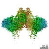 0905C  0906C 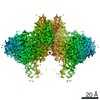 0907C 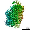 0908C  6lidC M: map data used to model this data C: citing same article ( |
|---|---|
| Similar structure data |


-Links
 PDBj
PDBj


- Assembly
| Deposited unit | 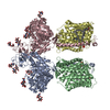
|
|---|---|
| 1 |
|
-Components
-Protein , 2 types, 4 molecules ACBD
| #1: Protein | Mass: 80691.586 Da / Num. of mol.: 2 Source method: isolated from a genetically manipulated source Source: (gene. exp.) Homo sapiens (human) / Gene: SLC3A1, RBAT / Production host: Homo sapiens (human) / References: UniProt: Q07837#2: Protein | Mass: 55656.105 Da / Num. of mol.: 2 Source method: isolated from a genetically manipulated source Source: (gene. exp.) Homo sapiens (human) / Gene: SLC7A9, BAT1 / Production host: Homo sapiens (human) / References: UniProt: P82251 |
|---|
-Sugars , 1 types, 10 molecules
| #3: Polysaccharide | 2-acetamido-2-deoxy-beta-D-glucopyranose-(1-4)-2-acetamido-2-deoxy-beta-D-glucopyranose Source method: isolated from a genetically manipulated source |
|---|
-Non-polymers , 4 types, 254 molecules 


| #4: Chemical |  Mass: 40.078 Da / Num. of mol.: 2 / Source method: obtained synthetically / Formula: Ca Mass: 40.078 Da / Num. of mol.: 2 / Source method: obtained synthetically / Formula: Ca#5: Chemical |  Type: L-peptide linking / Mass: 175.209 Da / Num. of mol.: 2 / Source method: obtained synthetically / Formula: C6H15N4O2 / Feature type: SUBJECT OF INVESTIGATION Type: L-peptide linking / Mass: 175.209 Da / Num. of mol.: 2 / Source method: obtained synthetically / Formula: C6H15N4O2 / Feature type: SUBJECT OF INVESTIGATION#6: Chemical | ChemComp-3PH /  Mass: 704.998 Da / Num. of mol.: 4 / Source method: obtained synthetically / Formula: C39H77O8P Mass: 704.998 Da / Num. of mol.: 4 / Source method: obtained synthetically / Formula: C39H77O8P#7: Water | ChemComp-HOH / | Mass: 18.015 Da / Num. of mol.: 246 / Source method: isolated from a natural source / Formula: H2O |
|---|
-Details
| Has ligand of interest | Y |
|---|---|
| Has protein modification | Y |
-Experimental details
-Experiment
| Experiment | Method: ELECTRON MICROSCOPY |
|---|---|
| EM experiment | Aggregation state: PARTICLE / 3D reconstruction method: single particle reconstruction |
- Sample preparation
| Component | Name: b0,+AT-rBAT complex bound with Arginine / Type: COMPLEX / Entity ID: #1-#2 / Source: RECOMBINANT | ||||||||||||||||
|---|---|---|---|---|---|---|---|---|---|---|---|---|---|---|---|---|---|
| Molecular weight |
| ||||||||||||||||
| Source (natural) | Organism: Homo sapiens (human) | ||||||||||||||||
| Source (recombinant) | Organism: Homo sapiens (human) | ||||||||||||||||
| Buffer solution | pH: 8 | ||||||||||||||||
| Specimen | Embedding applied: NO / Shadowing applied: NO / Staining applied: NO / Vitrification applied: YES | ||||||||||||||||
| Vitrification | Cryogen name: NITROGEN |
- Electron microscopy imaging
| Experimental equipment |  Model: Titan Krios / Image courtesy: FEI Company |
|---|---|
| Microscopy | Model: FEI TITAN KRIOS |
| Electron gun | Electron source:  FIELD EMISSION GUN / Accelerating voltage: 300 kV / Illumination mode: FLOOD BEAM FIELD EMISSION GUN / Accelerating voltage: 300 kV / Illumination mode: FLOOD BEAM |
| Electron lens | Mode: BRIGHT FIELD / Alignment procedure: COMA FREE |
| Specimen holder | Cryogen: NITROGEN / Specimen holder model: FEI TITAN KRIOS AUTOGRID HOLDER |
| Image recording | Electron dose: 50 e/Å2 / Film or detector model: GATAN K2 QUANTUM (4k x 4k) |
- Processing
| EM software |
| ||||||||||||
|---|---|---|---|---|---|---|---|---|---|---|---|---|---|
| CTF correction | Type: PHASE FLIPPING AND AMPLITUDE CORRECTION | ||||||||||||
| Symmetry | Point symmetry: C2 (2 fold cyclic) | ||||||||||||
| 3D reconstruction | Resolution: 2.3 Å / Resolution method: FSC 0.143 CUT-OFF / Num. of particles: 665827 / Symmetry type: POINT |
