 Movie
Movie Controller

[English] 日本語
 Yorodumi
- PDB-6lcm: Crystal structure of chloroplast resolvase ZmMOC1 with the magic ... -
+ Open data

- Basic information
| Entry | Database: PDB / ID: 6lcm | ||||||
|---|---|---|---|---|---|---|---|
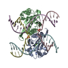
| Title | Crystal structure of chloroplast resolvase ZmMOC1 with the magic triangle I3C | ||||||
 Components Components | ZmMoc1 | ||||||
 Keywords Keywords | HYDROLASE / chloroplast / resolvase / Holliday junction / PLANT PROTEIN | ||||||
| Function / homology | Holliday junction resolvase MOC1-like / crossover junction DNA endonuclease activity / metal ion binding / Chem-I3C / Holliday junction resolvase MOC1, chloroplastic Function and homology information Function and homology information | ||||||
| Biological species |  | ||||||
| Method |  X-RAY DIFFRACTION / SAD / Resolution: 2.5 Å X-RAY DIFFRACTION / SAD / Resolution: 2.5 Å | ||||||
 Authors Authors | Yan, J.J. / Hong, S.X. / Guan, Z.Y. / Yin, P. | ||||||
 Citation Citation | Journal: Nat Commun / Year: 2020 Title: Structural insights into sequence-dependent Holliday junction resolution by the chloroplast resolvase MOC1. Authors: Yan, J. / Hong, S. / Guan, Z. / He, W. / Zhang, D. / Yin, P. | ||||||
| History |
|
- Structure visualization
| Structure viewer | Molecule: MolmilJmol/JSmol |
|---|
- Downloads & links
-Download
| PDBx/mmCIF format | 6lcm.cif.gz | 82.4 KB | Display | PDBx/mmCIF format |
|---|---|---|---|---|
| PDB format | pdb6lcm.ent.gz | 58.7 KB | Display | PDB format |
| PDBx/mmJSON format | 6lcm.json.gz | Tree view | PDBx/mmJSON format | |
| Others |  Other downloads Other downloads |
-Validation report
| Arichive directory | https://data.pdbj.org/pub/pdb/validation_reports/lc/6lcmftp://data.pdbj.org/pub/pdb/validation_reports/lc/6lcm | HTTPS FTP |
|---|
-Related structure data


-Links
 PDBj
PDBj- Assembly
| Deposited unit | 
| ||||||||||||
|---|---|---|---|---|---|---|---|---|---|---|---|---|---|
| 1 | 
| ||||||||||||
| Unit cell |
| ||||||||||||
| Components on special symmetry positions |
|
-Components
| #1: Protein | Mass: 19082.768 Da / Num. of mol.: 1 / Mutation: E158Q,I211V Source method: isolated from a genetically manipulated source Source: (gene. exp.)  | ||||
|---|---|---|---|---|---|
| #2: Chemical |   Mass: 558.835 Da / Num. of mol.: 3 / Source method: obtained synthetically / Formula: C8H4I3NO4 / Feature type: SUBJECT OF INVESTIGATION Mass: 558.835 Da / Num. of mol.: 3 / Source method: obtained synthetically / Formula: C8H4I3NO4 / Feature type: SUBJECT OF INVESTIGATION#3: Water | ChemComp-HOH / |  Mass: 18.015 Da / Num. of mol.: 53 / Source method: isolated from a natural source / Formula: H2O Mass: 18.015 Da / Num. of mol.: 53 / Source method: isolated from a natural source / Formula: H2OHas ligand of interest | Y | |
-Experimental details
-Experiment
| Experiment | Method: X-RAY DIFFRACTION / Number of used crystals: 1 |
|---|
- Sample preparation
| Crystal | Density Matthews: 2.24 Å3/Da / Density % sol: 45.16 % |
|---|---|
| Crystal grow | Temperature: 291.15 K / Method: vapor diffusion, hanging drop / Details: PEG 3350, Ammonium fluoride |
-Data collection
| Diffraction | Mean temperature: 100 K / Serial crystal experiment: N |
|---|---|
| Diffraction source | Source: ROTATING ANODE / Type: RIGAKU MICROMAX-007 HF / Wavelength: 1.5418 Å |
| Detector | Type: DECTRIS PILATUS3 R 200K-A / Detector: PIXEL / Date: Sep 2, 2017 |
| Radiation | Protocol: SINGLE WAVELENGTH / Monochromatic (M) / Laue (L): M / Scattering type: x-ray |
| Radiation wavelength | Wavelength: 1.5418 Å / Relative weight: 1 |
| Reflection | Resolution: 2.5→45 Å / Num. obs: 6571 / % possible obs: 100 % / Redundancy: 33.8 % / Rmerge(I) obs: 0.092 / Net I/σ(I): 70.8 |
| Reflection shell | Resolution: 2.5→2.59 Å / Rmerge(I) obs: 0.302 / Num. unique obs: 670 |
- Processing
| Software |
| ||||||||||||||||||||||||
|---|---|---|---|---|---|---|---|---|---|---|---|---|---|---|---|---|---|---|---|---|---|---|---|---|---|
| Refinement | Method to determine structure: SAD / Resolution: 2.5→45 Å / Cross valid method: FREE R-VALUE
| ||||||||||||||||||||||||
| Displacement parameters | Biso mean: 29.72 Å2 | ||||||||||||||||||||||||
| Refinement step | Cycle: LAST / Resolution: 2.5→45 Å
| ||||||||||||||||||||||||
| Refine LS restraints |
|
