 Movie
Movie Controller

[English] 日本語
 Yorodumi
- PDB-6l26: Neutron crystal structure of the mutant green fluorescent protein... -
+ Open data

- Basic information
| Entry | Database: PDB / ID: 6l26 | ||||||||||||
|---|---|---|---|---|---|---|---|---|---|---|---|---|---|
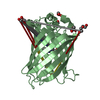
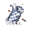
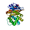
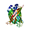
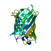
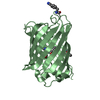
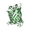
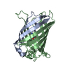
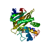
| Title | Neutron crystal structure of the mutant green fluorescent protein (EGFP) | ||||||||||||
 Components Components | Green fluorescent protein | ||||||||||||
 Keywords Keywords | FLUORESCENT PROTEIN / GFP / mutant / recombinant | ||||||||||||
| Function / homology | Green fluorescent protein, GFP / Green fluorescent protein-related / Green fluorescent protein / Green fluorescent protein / bioluminescence / generation of precursor metabolites and energy / trideuteriooxidanium / DEUTERATED WATER / Green fluorescent protein Function and homology information Function and homology information | ||||||||||||
| Biological species |   Aequorea victoria (jellyfish) Aequorea victoria (jellyfish) | ||||||||||||
| Method | NEUTRON DIFFRACTION / NUCLEAR REACTOR /  FOURIER SYNTHESIS / Resolution: 1.444 Å FOURIER SYNTHESIS / Resolution: 1.444 Å | ||||||||||||
 Authors Authors | Adachi, M. / Shimizu, R. / Shibazaki, C. / Kagotani, Y. / Ostermann, A. / Schrader, T.E. | ||||||||||||
| Funding support |  Japan, 1items Japan, 1items
| ||||||||||||
 Citation Citation | Journal: J Phys Chem Lett / Year: 2020 Title: Direct Observation of the Protonation States in the Mutant Green Fluorescent Protein. Authors: Shibazaki, C. / Shimizu, R. / Kagotani, Y. / Ostermann, A. / Schrader, T.E. / Adachi, M. | ||||||||||||
| History |
|
- Structure visualization
| Structure viewer | Molecule: MolmilJmol/JSmol |
|---|
- Downloads & links
-Download
| PDBx/mmCIF format | 6l26.cif.gz | 127.4 KB | Display | PDBx/mmCIF format |
|---|---|---|---|---|
| PDB format | pdb6l26.ent.gz | 101.8 KB | Display | PDB format |
| PDBx/mmJSON format | 6l26.json.gz | Tree view | PDBx/mmJSON format | |
| Others |  Other downloads Other downloads |
-Validation report
| Arichive directory | https://data.pdbj.org/pub/pdb/validation_reports/l2/6l26ftp://data.pdbj.org/pub/pdb/validation_reports/l2/6l26 | HTTPS FTP |
|---|
-Related structure data
| Related structure data |  6l27C 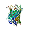 2wurS S: Starting model for refinement C: citing same article ( |
|---|---|
| Similar structure data |

-Links
 PDBj
PDBj


- Assembly
| Deposited unit | 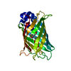
| ||||||||
|---|---|---|---|---|---|---|---|---|---|
| 1 |
| ||||||||
| Unit cell |
|
-Components
| #1: Protein | Mass: 25843.053 Da / Num. of mol.: 1 Source method: isolated from a genetically manipulated source Details: The amino acids of TYG are processed to chromophore as CRO by post-translational reaction. Source: (gene. exp.) Aequorea victoria (jellyfish) / Gene: gfp / Production host:  |
|---|---|
| #2: Chemical | ChemComp-D3O /   Mass: 22.042 Da / Num. of mol.: 1 / Source method: isolated from a natural source / Formula: D3O / Feature type: SUBJECT OF INVESTIGATION Mass: 22.042 Da / Num. of mol.: 1 / Source method: isolated from a natural source / Formula: D3O / Feature type: SUBJECT OF INVESTIGATION |
| #3: Chemical | ChemComp-DOD /   Mass: 18.015 Da / Num. of mol.: 372 / Source method: isolated from a natural source / Formula: D2O Mass: 18.015 Da / Num. of mol.: 372 / Source method: isolated from a natural source / Formula: D2O |
| Has ligand of interest | Y |
| Has protein modification | Y |
| Sequence details | Residue SER 66 has been mutated to THR 66. Residues THR 66, TYR 67 and GLY 68 constitute the ...Residue SER 66 has been mutated to THR 66. Residues THR 66, TYR 67 and GLY 68 constitute the chromophore CRO 66. |
-Experimental details
-Experiment
| Experiment | Method: NEUTRON DIFFRACTION / Number of used crystals: 1 |
|---|
- Sample preparation
| Crystal | Density Matthews: 2.12 Å3/Da / Density % sol: 41.96 % |
|---|---|
| Crystal grow | Temperature: 293 K / Method: vapor diffusion, sitting drop Details: 0.1 M MES-NaOD (pD 7.0), 5.0 % w/v PEG 2000 and 50 mM NDSB |
-Data collection
| Diffraction | Mean temperature: 100 K / Serial crystal experiment: N |
|---|---|
| Diffraction source | Source: NUCLEAR REACTOR / Site: FRM II  / Beamline: BIODIFF / Wavelength: 2.668 Å / Beamline: BIODIFF / Wavelength: 2.668 Å |
| Detector | Type: BIODIFF / Detector: IMAGE PLATE / Date: Nov 14, 2017 |
| Radiation | Protocol: SINGLE WAVELENGTH / Monochromatic (M) / Laue (L): M / Scattering type: neutron |
| Radiation wavelength | Wavelength: 2.668 Å / Relative weight: 1 |
| Reflection | Resolution: 1.45→24.8 Å / Num. obs: 90310 / % possible obs: 88.9 % / Redundancy: 2.5 % / Rmerge(I) obs: 0.098 / Net I/σ(I): 7.9 |
| Reflection shell | Resolution: 1.45→1.49 Å / Redundancy: 1.9 % / Rmerge(I) obs: 0.177 / Mean I/σ(I) obs: 1.9 / Num. unique obs: 25594 / % possible all: 73.8 |
- Processing
| Software |
| ||||||||||||||||||||||||||||||||||||||||||||||||||||||||||||||||||||||||||||||||||||
|---|---|---|---|---|---|---|---|---|---|---|---|---|---|---|---|---|---|---|---|---|---|---|---|---|---|---|---|---|---|---|---|---|---|---|---|---|---|---|---|---|---|---|---|---|---|---|---|---|---|---|---|---|---|---|---|---|---|---|---|---|---|---|---|---|---|---|---|---|---|---|---|---|---|---|---|---|---|---|---|---|---|---|---|---|---|
| Refinement | Method to determine structure: FOURIER SYNTHESIS Starting model: 2WUR Resolution: 1.444→24.764 Å / SU ML: 0.07 / Cross valid method: FREE R-VALUE / σ(F): 0 / Phase error: 15.37
| ||||||||||||||||||||||||||||||||||||||||||||||||||||||||||||||||||||||||||||||||||||
| Solvent computation | Shrinkage radii: 0.9 Å / VDW probe radii: 1.11 Å | ||||||||||||||||||||||||||||||||||||||||||||||||||||||||||||||||||||||||||||||||||||
| Refinement step | Cycle: final / Resolution: 0.88→39.362 Å
| ||||||||||||||||||||||||||||||||||||||||||||||||||||||||||||||||||||||||||||||||||||
| Refine LS restraints |
| ||||||||||||||||||||||||||||||||||||||||||||||||||||||||||||||||||||||||||||||||||||
| LS refinement shell | Refine-ID: NEUTRON DIFFRACTION / Rfactor Rfree error: 0
|
