 Movie
Movie Controller


+ Open data

- Basic information
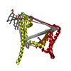
| Entry | Database: PDB / ID: 6kzp | ||||||||||||||||||
|---|---|---|---|---|---|---|---|---|---|---|---|---|---|---|---|---|---|---|---|
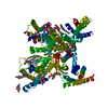
| Title | calcium channel-ligand | ||||||||||||||||||
 Components Components | Voltage-dependent T-type calcium channel subunit alpha-1G,Voltage-dependent T-type calcium channel subunit alpha-1G | ||||||||||||||||||
 Keywords Keywords | MEMBRANE PROTEIN / channel / blocker | ||||||||||||||||||
| Function / homology |  Function and homology information Function and homology informationSA node cell to atrial cardiac muscle cell signaling / AV node cell to bundle of His cell signaling / voltage-gated calcium channel activity involved SA node cell action potential / sinoatrial node development / low voltage-gated calcium channel activity / response to nickel cation / voltage-gated calcium channel activity involved in AV node cell action potential / AV node cell action potential / SA node cell action potential / membrane depolarization during SA node cell action potential ...SA node cell to atrial cardiac muscle cell signaling / AV node cell to bundle of His cell signaling / voltage-gated calcium channel activity involved SA node cell action potential / sinoatrial node development / low voltage-gated calcium channel activity / response to nickel cation / voltage-gated calcium channel activity involved in AV node cell action potential / AV node cell action potential / SA node cell action potential / membrane depolarization during SA node cell action potential / high voltage-gated calcium channel activity / regulation of atrial cardiac muscle cell membrane depolarization / membrane depolarization during AV node cell action potential / calcium ion import / NCAM1 interactions / cardiac muscle cell action potential involved in contraction / voltage-gated calcium channel complex / regulation of heart rate by cardiac conduction / calcium ion import across plasma membrane / Smooth Muscle Contraction / voltage-gated calcium channel activity / regulation of membrane potential / calcium ion transmembrane transport / scaffold protein binding / chemical synaptic transmission / synapse / plasma membrane / cytoplasm Similarity search - Function | ||||||||||||||||||
| Biological species |  Homo sapiens (human) Homo sapiens (human) | ||||||||||||||||||
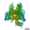
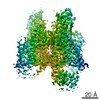
| Method | ELECTRON MICROSCOPY / single particle reconstruction / cryo EM / Resolution: 3.1 Å | ||||||||||||||||||
 Authors Authors | Yan, N. | ||||||||||||||||||
| Funding support |  China, 5items China, 5items
| ||||||||||||||||||
 Citation Citation | Journal: Nature / Year: 2019 Title: Cryo-EM structures of apo and antagonist-bound human Ca3.1. Authors: Yanyu Zhao / Gaoxingyu Huang / Qiurong Wu / Kun Wu / Ruiqi Li / Jianlin Lei / Xiaojing Pan / Nieng Yan /  Abstract: Among the ten subtypes of mammalian voltage-gated calcium (Ca) channels, Ca3.1-Ca3.3 constitute the T-type, or the low-voltage-activated, subfamily, the abnormal activities of which are associated ...Among the ten subtypes of mammalian voltage-gated calcium (Ca) channels, Ca3.1-Ca3.3 constitute the T-type, or the low-voltage-activated, subfamily, the abnormal activities of which are associated with epilepsy, psychiatric disorders and pain. Here we report the cryo-electron microscopy structures of human Ca3.1 alone and in complex with a highly Ca3-selective blocker, Z944, at resolutions of 3.3 Å and 3.1 Å, respectively. The arch-shaped Z944 molecule reclines in the central cavity of the pore domain, with the wide end inserting into the fenestration on the interface between repeats II and III, and the narrow end hanging above the intracellular gate like a plug. The structures provide the framework for comparative investigation of the distinct channel properties of different Ca subfamilies. | ||||||||||||||||||
| History |
|
- Structure visualization
| Movie |
Movie viewer |
|---|---|
| Structure viewer | Molecule: MolmilJmol/JSmol |
- Downloads & links
-Download
| PDBx/mmCIF format | 6kzp.cif.gz | 232 KB | Display | PDBx/mmCIF format |
|---|---|---|---|---|
| PDB format | pdb6kzp.ent.gz | 164.1 KB | Display | PDB format |
| PDBx/mmJSON format | 6kzp.json.gz | Tree view | PDBx/mmJSON format | |
| Others |  Other downloads Other downloads |
-Validation report
| Arichive directory | https://data.pdbj.org/pub/pdb/validation_reports/kz/6kzpftp://data.pdbj.org/pub/pdb/validation_reports/kz/6kzp | HTTPS FTP |
|---|
-Related structure data
| Related structure data |  0792MC  0791C 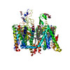 6kzoC M: map data used to model this data C: citing same article ( |
|---|---|
| Similar structure data |


-Links
 PDBj
PDBj


- Assembly
| Deposited unit | 
|
|---|---|
| 1 |
|
-Components
-Protein / Sugars , 2 types, 5 molecules A
| #1: Protein | Mass: 238874.984 Da / Num. of mol.: 1 Source method: isolated from a genetically manipulated source Source: (gene. exp.) Homo sapiens (human) / Gene: CACNA1G, KIAA1123 / Production host: Homo sapiens (human) / References: UniProt: O43497 |
|---|---|
| #3: Sugar | ChemComp-NAG /  Type: D-saccharide, beta linking / Mass: 221.208 Da / Num. of mol.: 4 Type: D-saccharide, beta linking / Mass: 221.208 Da / Num. of mol.: 4Source method: isolated from a genetically manipulated source Formula: C8H15NO6 |
-Non-polymers , 4 types, 14 molecules 


| #2: Chemical |  Mass: 40.078 Da / Num. of mol.: 2 / Source method: obtained synthetically / Formula: Ca Mass: 40.078 Da / Num. of mol.: 2 / Source method: obtained synthetically / Formula: Ca#4: Chemical | ChemComp-DZR / ~{ |  Mass: 383.888 Da / Num. of mol.: 1 / Source method: obtained synthetically / Formula: C19H27ClFN3O2 / Feature type: SUBJECT OF INVESTIGATION / Comment: Z944*YM Mass: 383.888 Da / Num. of mol.: 1 / Source method: obtained synthetically / Formula: C19H27ClFN3O2 / Feature type: SUBJECT OF INVESTIGATION / Comment: Z944*YM#5: Chemical | ChemComp-3PE /  Mass: 748.065 Da / Num. of mol.: 8 / Source method: obtained synthetically / Formula: C41H82NO8P / Comment: phospholipid*YM Mass: 748.065 Da / Num. of mol.: 8 / Source method: obtained synthetically / Formula: C41H82NO8P / Comment: phospholipid*YM#6: Chemical |  Mass: 486.726 Da / Num. of mol.: 3 / Source method: obtained synthetically / Formula: C31H50O4 Mass: 486.726 Da / Num. of mol.: 3 / Source method: obtained synthetically / Formula: C31H50O4 |
|---|
-Details
| Has ligand of interest | Y |
|---|---|
| Has protein modification | Y |
-Experimental details
-Experiment
| Experiment | Method: ELECTRON MICROSCOPY |
|---|---|
| EM experiment | Aggregation state: PARTICLE / 3D reconstruction method: single particle reconstruction |
- Sample preparation
| Component | Name: membrane protein-ligand / Type: COMPLEX / Entity ID: #1 / Source: RECOMBINANT |
|---|---|
| Source (natural) | Organism: Homo sapiens (human) |
| Source (recombinant) | Organism: Homo sapiens (human) |
| Buffer solution | pH: 7.4 |
| Specimen | Embedding applied: NO / Shadowing applied: NO / Staining applied: NO / Vitrification applied: YES |
| Vitrification | Cryogen name: ETHANE |
- Electron microscopy imaging
| Experimental equipment |  Model: Titan Krios / Image courtesy: FEI Company |
|---|---|
| Microscopy | Model: FEI TITAN KRIOS |
| Electron gun | Electron source:  FIELD EMISSION GUN / Accelerating voltage: 300 kV / Illumination mode: FLOOD BEAM FIELD EMISSION GUN / Accelerating voltage: 300 kV / Illumination mode: FLOOD BEAM |
| Electron lens | Mode: BRIGHT FIELD |
| Image recording | Electron dose: 48 e/Å2 / Film or detector model: GATAN K2 SUMMIT (4k x 4k) |
- Processing
| CTF correction | Type: PHASE FLIPPING ONLY |
|---|---|
| 3D reconstruction | Resolution: 3.1 Å / Resolution method: FSC 0.143 CUT-OFF / Num. of particles: 138449 / Symmetry type: POINT |
