 Movie
Movie Controller


+ Open data

- Basic information
| Entry | Database: EMDB / ID: EMD-0792 | ||||||||||||||||||
|---|---|---|---|---|---|---|---|---|---|---|---|---|---|---|---|---|---|---|---|
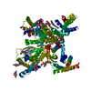
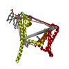
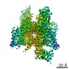
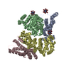
| Title | calcium channel-ligand | ||||||||||||||||||
 Map data Map data | |||||||||||||||||||
 Sample Sample |
| ||||||||||||||||||
 Keywords Keywords | channel / blocker / MEMBRANE PROTEIN | ||||||||||||||||||
| Function / homology |  Function and homology information Function and homology informationSA node cell to atrial cardiac muscle cell signaling / AV node cell to bundle of His cell signaling / voltage-gated calcium channel activity involved SA node cell action potential / sinoatrial node development / low voltage-gated calcium channel activity / response to nickel cation / voltage-gated calcium channel activity involved in AV node cell action potential / AV node cell action potential / SA node cell action potential / membrane depolarization during SA node cell action potential ...SA node cell to atrial cardiac muscle cell signaling / AV node cell to bundle of His cell signaling / voltage-gated calcium channel activity involved SA node cell action potential / sinoatrial node development / low voltage-gated calcium channel activity / response to nickel cation / voltage-gated calcium channel activity involved in AV node cell action potential / AV node cell action potential / SA node cell action potential / membrane depolarization during SA node cell action potential / high voltage-gated calcium channel activity / regulation of atrial cardiac muscle cell membrane depolarization / membrane depolarization during AV node cell action potential / calcium ion import / NCAM1 interactions / cardiac muscle cell action potential involved in contraction / voltage-gated calcium channel complex / regulation of heart rate by cardiac conduction / calcium ion import across plasma membrane / Smooth Muscle Contraction / voltage-gated calcium channel activity / regulation of membrane potential / calcium ion transmembrane transport / scaffold protein binding / chemical synaptic transmission / synapse / plasma membrane / cytoplasm Similarity search - Function | ||||||||||||||||||
| Biological species |  Homo sapiens (human) Homo sapiens (human) | ||||||||||||||||||
| Method | single particle reconstruction / cryo EM / Resolution: 3.1 Å | ||||||||||||||||||
 Authors Authors | Yan N | ||||||||||||||||||
| Funding support |  China, 5 items China, 5 items
| ||||||||||||||||||
 Citation Citation | Journal: Nature / Year: 2019 Title: Cryo-EM structures of apo and antagonist-bound human Ca3.1. Authors: Yanyu Zhao / Gaoxingyu Huang / Qiurong Wu / Kun Wu / Ruiqi Li / Jianlin Lei / Xiaojing Pan / Nieng Yan /  Abstract: Among the ten subtypes of mammalian voltage-gated calcium (Ca) channels, Ca3.1-Ca3.3 constitute the T-type, or the low-voltage-activated, subfamily, the abnormal activities of which are associated ...Among the ten subtypes of mammalian voltage-gated calcium (Ca) channels, Ca3.1-Ca3.3 constitute the T-type, or the low-voltage-activated, subfamily, the abnormal activities of which are associated with epilepsy, psychiatric disorders and pain. Here we report the cryo-electron microscopy structures of human Ca3.1 alone and in complex with a highly Ca3-selective blocker, Z944, at resolutions of 3.3 Å and 3.1 Å, respectively. The arch-shaped Z944 molecule reclines in the central cavity of the pore domain, with the wide end inserting into the fenestration on the interface between repeats II and III, and the narrow end hanging above the intracellular gate like a plug. The structures provide the framework for comparative investigation of the distinct channel properties of different Ca subfamilies. | ||||||||||||||||||
| History |
|
- Structure visualization
| Movie |
Movie viewer |
|---|---|
| Structure viewer | EM map: SurfViewMolmilJmol/JSmol |
| Supplemental images |

- Downloads & links
-EMDB archive
| Map data | emd_0792.map.gz | 116.6 MB | EMDB map data format | |
|---|---|---|---|---|
| Header (meta data) | emd-0792-v30.xmlemd-0792.xml | 13.8 KB 13.8 KB | Display Display | EMDB header |
| Images |  emd_0792.png emd_0792.png | 175.1 KB | ||
| Masks | emd_0792_msk_1.map | 125 MB | Mask map | |
| Filedesc metadata | emd-0792.cif.gz | 6.7 KB | ||
| Archive directory |  http://ftp.pdbj.org/pub/emdb/structures/EMD-0792ftp://ftp.pdbj.org/pub/emdb/structures/EMD-0792 http://ftp.pdbj.org/pub/emdb/structures/EMD-0792ftp://ftp.pdbj.org/pub/emdb/structures/EMD-0792 | HTTPS FTP |
-Related structure data
| Related structure data |  6kzpMC  0791C 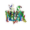 6kzoC M: atomic model generated by this map C: citing same article ( |
|---|---|
| Similar structure data |


-Links
| EMDB pages | EMDB (EBI/PDBe) / EMDataResource |
|---|---|
| Related items in Molecule of the Month |


-Map
| File | Download / File: emd_0792.map.gz / Format: CCP4 / Size: 125 MB / Type: IMAGE STORED AS FLOATING POINT NUMBER (4 BYTES) | ||||||||||||||||||||||||||||||||||||||||||||||||||||||||||||||||||||
|---|---|---|---|---|---|---|---|---|---|---|---|---|---|---|---|---|---|---|---|---|---|---|---|---|---|---|---|---|---|---|---|---|---|---|---|---|---|---|---|---|---|---|---|---|---|---|---|---|---|---|---|---|---|---|---|---|---|---|---|---|---|---|---|---|---|---|---|---|---|
| Projections & slices | Image control
Images are generated by Spider. | ||||||||||||||||||||||||||||||||||||||||||||||||||||||||||||||||||||
| Voxel size | X=Y=Z: 1.091 Å | ||||||||||||||||||||||||||||||||||||||||||||||||||||||||||||||||||||
| Density |
| ||||||||||||||||||||||||||||||||||||||||||||||||||||||||||||||||||||
| Symmetry | Space group: 1 | ||||||||||||||||||||||||||||||||||||||||||||||||||||||||||||||||||||
| Details | EMDB XML:
CCP4 map header:
| ||||||||||||||||||||||||||||||||||||||||||||||||||||||||||||||||||||
 Z (Sec.)
 Y (Row.)
 X (Col.)


-Supplemental data
-Mask #1
| File | emd_0792_msk_1.map | ||||||||||||
|---|---|---|---|---|---|---|---|---|---|---|---|---|---|
| Projections & Slices |
| ||||||||||||
| Density Histograms |


- Sample components
-Entire : membrane protein-ligand
| Entire | Name: membrane protein-ligand |
|---|---|
| Components |
|
-Supramolecule #1: membrane protein-ligand
| Supramolecule | Name: membrane protein-ligand / type: complex / ID: 1 / Parent: 0 / Macromolecule list: #1 |
|---|---|
| Source (natural) | Organism: Homo sapiens (human) |
-Macromolecule #1: Voltage-dependent T-type calcium channel subunit alpha-1G,Voltage...
| Macromolecule | Name: Voltage-dependent T-type calcium channel subunit alpha-1G,Voltage-dependent T-type calcium channel subunit alpha-1G type: protein_or_peptide / ID: 1 / Number of copies: 1 / Enantiomer: LEVO |
|---|---|
| Source (natural) | Organism: Homo sapiens (human) |
| Molecular weight | Theoretical: 238.874984 KDa |
| Recombinant expression | Organism: Homo sapiens (human) |
| Sequence | String: MHHHHHHHHG DYKDDDDKGT DEEEDGAGAE ESGQPRSFMR LNDLSGAGGR PGPGSAEKDP GSADSEAEGL PYPALAPVVF FYLSQDSRP RSWCLRTVCN PWFERISMLV ILLNCVTLGM FRPCEDIACD SQRCRILQAF DDFIFAFFAV EMVVKMVALG I FGKKCYLG ...String: MHHHHHHHHG DYKDDDDKGT DEEEDGAGAE ESGQPRSFMR LNDLSGAGGR PGPGSAEKDP GSADSEAEGL PYPALAPVVF FYLSQDSRP RSWCLRTVCN PWFERISMLV ILLNCVTLGM FRPCEDIACD SQRCRILQAF DDFIFAFFAV EMVVKMVALG I FGKKCYLG DTWNRLDFFI VIAGMLEYSL DLQNVSFSAV RTVRVLRPLR AINRVPSMRI LVTLLLDTLP MLGNVLLLCF FV FFIFGIV GVQLWAGLLR NRCFLPENFS LPLSVDLERY YQTENEDESP FICSQPRENG MRSCRSVPTL RGDGGGGPPC GLD YEAYNS SSNTTCVNWN QYYTNCSAGE HNPFKGAINF DNIGYAWIAI FQVITLEGWV DIMYFVMDAH SFYNFIYFIL LIIV GSFFM INLCLVVIAT QFSETKQRES QLMREQRVRF LSNASTLASF SEPGSCYEEL LKYLVYILRK AARRLAQVSR AAGVR VGLL SSPAPLGGQE TQPSSSCSRS HRRLSVHHLV HHHHHHHHHY HLGACQSSCK ISSPCLKADS GACGPDSCPY CARAGA GEV ELADREMPDS DSEAVYEFTQ DAQHSDLRDP HSRRQRSLGP DAEPSSVLAF WRLICDTFRK IVDSKYFGRG IMIAILV NT LSMGIEYHEQ PEELTNALEI SNIVFTSLFA LEMLLKLLVY GPFGYIKNPY NIFDGVIVVI SVWEIVGQQG GGLSVLRT F RLMRVLKLVR FLPALQRQLV VLMKTMDNVA TFCMLLMLFI FIFSILGMHL FGCKFASERD GDTLPDRKNF DSLLWAIVT VFQILTQEDW NKVLYNGMAS TSSWAALYFI ALMTFGNYVL FNLLVAILVE GFQAEGDANK SESEPDFFSP SLDGDGDRKK CLALVSLGE HPELRKSLLP PLIIHTAATP MSLPKSTSTG LGEALGPASR RTSSSGSAEP GAAHEMKSPP SARSSPHSPW S AASSWTSR RSSRNSLGRA PSLKRRSPSG ERRSLLSGEG QESQDEEESS EEERASPAGS DHRHRGSLER EAKSSFDLPD TL QVPGLHR TASGRGSASE HQDCNGKSAS GRLARALRPD DPPLDGDDAD DEGNLSKGER VRAWIRARLP ACCLERDSWS AYI FPPQSR FRLLCHRIIT HKMFDHVVLV IIFLNCITIA MERPKIDPHS AERIFLTLSN YIFTAVFLAE MTVKVVALGW CFGE QAYLR SSWNVLDGLL VLISVIDILV SMVSDSGTKI LGMLRVLRLL RTLRPLRVIS RAQGLKLVVE TLMSSLKPIG NIVVI CCAF FIIFGILGVQ LFKGKFFVCQ GEDTRNITNK SDCAEASYRW VRHKYNFDNL GQALMSLFVL ASKDGWVDIM YDGLDA VGV DQQPIMNHNP WMLLYFISFL LIVAFFVLNM FVGVVVENFH KCRQHQEEEE ARRREEKRLR RLEKKRRNLM LDDVIAS GS SASAASEAQC KPYYSDYSRF RLLVHHLCTS HYLDLFITGV IGLNVVTMAM EHYQQPQILD EALKICNYIF TVIFVLES V FKLVAFGFRR FFQDRWNQLD LAIVLLSIMG ITLEEIEVNA SLPINPTIIR IMRVLRIARV LKLLKMAVGM RALLDTVMQ ALPQVGNLGL LFMLLFFIFA ALGVELFGDL ECDETHPCEG LGRHATFRNF GMAFLTLFRV STGDNWNGIM KDTLRDCDQE STCYNTVIS PIYFVSFVLT AQFVLVNVVI AVLMKHLEES NKEAKEEAEL EAELELEMKT LSPQPHSPLG SPFLWPGVEG P DSPDSPKP GALHPAAHAR SASHFSLEHP TMQPHPTELP GPDLLTVRKS GVSRTHSLPN DSYMCRHGST AEGPLGHRGW GL PKAQSGS VLSVHSQPAD TSYILQLPKD APHLLQPHSA PTWGTIPKLP PPGRSPLAQR PLRRQAAIRT DSLDVQGLGS RED LLAEVS GPSPPLARAY SFWGQSSTQA QQHSRSHSKI SKHMTPPAPC PGPEPNWGKG PPETRSSLEL DTELSWISGD LLPP GGQEE PPSPRDLKKC YSVEAQSCQR RPTSWLDEQR RHSIAVSCLD SGSQPHLGTD PSNLGGQPLG GPGSRPKKKL SPPSI TIDP PESQGPRTPP SPGICLRRRA PSSDSKDPLA SGPPDSMAAS PSPKKDVLSL SGLSSDPADL DP UniProtKB: Voltage-dependent T-type calcium channel subunit alpha-1G, Voltage-dependent T-type calcium channel subunit alpha-1G |
-Macromolecule #2: CALCIUM ION
| Macromolecule | Name: CALCIUM ION / type: ligand / ID: 2 / Number of copies: 2 / Formula: CA |
|---|---|
| Molecular weight | Theoretical: 40.078 Da |
-Macromolecule #3: 2-acetamido-2-deoxy-beta-D-glucopyranose
| Macromolecule | Name: 2-acetamido-2-deoxy-beta-D-glucopyranose / type: ligand / ID: 3 / Number of copies: 4 / Formula: NAG |
|---|---|
| Molecular weight | Theoretical: 221.208 Da |
| Chemical component information |  ChemComp-NAG: |
-Macromolecule #4: ~{N}-[[1-[2-(~{tert}-butylamino)-2-oxidanylidene-ethyl]piperidin-...
| Macromolecule | Name: ~{N}-[[1-[2-(~{tert}-butylamino)-2-oxidanylidene-ethyl]piperidin-4-yl]methyl]-3-chloranyl-5-fluoranyl-benzamide type: ligand / ID: 4 / Number of copies: 1 / Formula: DZR |
|---|---|
| Molecular weight | Theoretical: 383.888 Da |
| Chemical component information |  ChemComp-DZR: |
-Macromolecule #5: 1,2-Distearoyl-sn-glycerophosphoethanolamine
| Macromolecule | Name: 1,2-Distearoyl-sn-glycerophosphoethanolamine / type: ligand / ID: 5 / Number of copies: 8 / Formula: 3PE |
|---|---|
| Molecular weight | Theoretical: 748.065 Da |
| Chemical component information |  ChemComp-3PE: |
-Macromolecule #6: CHOLESTEROL HEMISUCCINATE
| Macromolecule | Name: CHOLESTEROL HEMISUCCINATE / type: ligand / ID: 6 / Number of copies: 3 / Formula: Y01 |
|---|---|
| Molecular weight | Theoretical: 486.726 Da |
| Chemical component information |  ChemComp-Y01: |
-Experimental details
-Structure determination
| Method | cryo EM |
|---|---|
 Processing Processing | single particle reconstruction |
| Aggregation state | particle |
-Sample preparation
| Buffer | pH: 7.4 |
|---|---|
| Vitrification | Cryogen name: ETHANE |
- Electron microscopy
| Microscope | FEI TITAN KRIOS |
|---|---|
| Image recording | Film or detector model: GATAN K2 SUMMIT (4k x 4k) / Average electron dose: 48.0 e/Å2 |
| Electron beam | Acceleration voltage: 300 kV / Electron source:  FIELD EMISSION GUN FIELD EMISSION GUN |
| Electron optics | Illumination mode: FLOOD BEAM / Imaging mode: BRIGHT FIELD |
| Experimental equipment |  Model: Titan Krios / Image courtesy: FEI Company |
-Image processing
| Startup model | Type of model: EMDB MAP |
|---|---|
| Final reconstruction | Resolution.type: BY AUTHOR / Resolution: 3.1 Å / Resolution method: FSC 0.143 CUT-OFF / Number images used: 138449 |
| Initial angle assignment | Type: MAXIMUM LIKELIHOOD |
| Final angle assignment | Type: MAXIMUM LIKELIHOOD |
