- PDB-6kzj: Crystal structure of Ankyrin B/NdeL1 complex -
+
Open data
ID or keywords:
Loading...
-
Basic information
Entry
Database: PDB / ID: 6kzj
Title
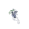
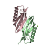
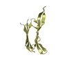
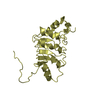
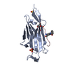
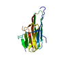
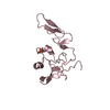
Crystal structure of Ankyrin B/NdeL1 complex
Components
Ankyrin-2
Nuclear distribution protein nudE-like 1
Keywords
PROTEIN BINDING / PROTEIN TRANSPORT / STRUCTURAL PROTEIN
Function / homology
Function and homology information
neurofilament cytoskeleton / protein localization to T-tubule / atrial cardiac muscle cell to AV node cell communication / SA node cell to atrial cardiac muscle cell communication / protein localization to M-band / T-tubule organization / central nervous system neuron axonogenesis / SA node cell action potential / membrane depolarization during SA node cell action potential / paranodal junction assembly ...neurofilament cytoskeleton / protein localization to T-tubule / atrial cardiac muscle cell to AV node cell communication / SA node cell to atrial cardiac muscle cell communication / protein localization to M-band / T-tubule organization / central nervous system neuron axonogenesis / SA node cell action potential / membrane depolarization during SA node cell action potential / paranodal junction assembly / oligopeptidase activity / Amplification of signal from unattached kinetochores via a MAD2 inhibitory signal / central region of growth cone / nuclear membrane disassembly / potassium channel activator activity / positive regulation of potassium ion import across plasma membrane / regulation of atrial cardiac muscle cell action potential / cerebral cortex radially oriented cell migration / Mitotic Prometaphase / EML4 and NUDC in mitotic spindle formation / Resolution of Sister Chromatid Cohesion / channel activator activity / phosphorylation-dependent protein binding / RHO GTPases Activate Formins / sarcoplasmic reticulum calcium ion transport / Separation of Sister Chromatids / regulation of SA node cell action potential / atrial cardiac muscle cell action potential / positive regulation of axon regeneration / protein localization to endoplasmic reticulum / A band / atrial septum development / neurofilament cytoskeleton organization / axon hillock / cytoskeletal adaptor activity / lysosome localization / costamere / microtubule nucleation / ventricular cardiac muscle cell action potential / regulation of ventricular cardiac muscle cell membrane repolarization / positive regulation of calcium ion transport / response to methylmercury / mitotic centrosome separation / regulation of release of sequestered calcium ion into cytosol / regulation of cardiac muscle cell contraction / vesicle transport along microtubule / protein localization to cell surface / retrograde axonal transport / M band / neuron projection extension / microtubule associated complex / centrosome localization / regulation of cardiac muscle contraction by calcium ion signaling / kinesin complex / Interaction between L1 and Ankyrins / inner cell mass cell proliferation / regulation of intracellular protein transport / positive regulation of ruffle assembly / spectrin binding / regulation of neuron projection development / establishment of mitotic spindle orientation / cell leading edge / regulation of calcium ion transport / microtubule organizing center / regulation of heart rate by cardiac conduction / intercalated disc / regulation of cardiac muscle contraction / alpha-tubulin binding / positive regulation of axon extension / beta-tubulin binding / COPI-mediated anterograde transport / regulation of cardiac muscle contraction by regulation of the release of sequestered calcium ion / axon cytoplasm / T-tubule / regulation of heart rate / protein localization to plasma membrane / chromosome segregation / regulation of protein stability / sarcolemma / recycling endosome / structural constituent of cytoskeleton / kinetochore / microtubule cytoskeleton organization / spindle / neuron migration / Z disc / endocytosis / intracellular calcium ion homeostasis / neuron projection development / synaptic vesicle / insulin receptor signaling pathway / intracellular protein localization / nuclear envelope / cell migration / protein transport / ATPase binding / cell body / microtubule binding / basolateral plasma membrane / cytoskeleton Similarity search - Function
NUDE domain / NUDE family / NUDE protein, C-terminal conserved region / Ankyrin, UPA domain / UPA domain / : / Domain present in ZO-1 and Unc5-like netrin receptors / ZU5 domain / ZU5 domain / ZU5 domain profile. ...NUDE domain / NUDE family / NUDE protein, C-terminal conserved region / Ankyrin, UPA domain / UPA domain / : / Domain present in ZO-1 and Unc5-like netrin receptors / ZU5 domain / ZU5 domain / ZU5 domain profile. / Death domain profile. / DEATH domain, found in proteins involved in cell death (apoptosis). / Death domain / Death domain / Ankyrin repeats (many copies) / Death-like domain superfamily / Ankyrin repeat / Ankyrin repeat profile. / Ankyrin repeats (3 copies) / Ankyrin repeat region circular profile. / ankyrin repeats / Ankyrin repeat / Ankyrin repeat-containing domain superfamily Similarity search - Domain/homology
Method to determine structure: AB INITIO PHASING / Resolution: 1.5→10 Å / Cor.coef. Fo:Fc: 0.966 / Cor.coef. Fo:Fc free: 0.953 / SU B: 2.842 / SU ML: 0.048 / SU R Cruickshank DPI: 0.0836 / Cross valid method: THROUGHOUT / σ(F): 0 / ESU R: 0.084 / ESU R Free: 0.077 Details: U VALUES : WITH TLS ADDED HYDROGENS HAVE BEEN ADDED IN THE RIDING POSITIONS
Rfactor
Num. reflection
% reflection
Selection details
Rfree
0.2108
1063
4.8 %
RANDOM
Rwork
0.1605
-
-
-
obs
0.1628
20976
94.53 %
-
Solvent computation
Ion probe radii: 0.8 Å / Shrinkage radii: 0.8 Å / VDW probe radii: 1.2 Å
In the structure databanks used in Yorodumi, some data are registered as the other names, "COVID-19 virus" and "2019-nCoV". Here are the details of the virus and the list of structure data.
Jan 31, 2019. EMDB accession codes are about to change! (news from PDBe EMDB page)
EMDB accession codes are about to change! (news from PDBe EMDB page)
The allocation of 4 digits for EMDB accession codes will soon come to an end. Whilst these codes will remain in use, new EMDB accession codes will include an additional digit and will expand incrementally as the available range of codes is exhausted. The current 4-digit format prefixed with “EMD-” (i.e. EMD-XXXX) will advance to a 5-digit format (i.e. EMD-XXXXX), and so on. It is currently estimated that the 4-digit codes will be depleted around Spring 2019, at which point the 5-digit format will come into force.
The EM Navigator/Yorodumi systems omit the EMD- prefix.
Related info.:Q: What is EMD? / ID/Accession-code notation in Yorodumi/EM Navigator
Yorodumi is a browser for structure data from EMDB, PDB, SASBDB, etc.
This page is also the successor to EM Navigator detail page, and also detail information page/front-end page for Omokage search.
The word "yorodu" (or yorozu) is an old Japanese word meaning "ten thousand". "mi" (miru) is to see.
Related info.:EMDB / PDB / SASBDB / Comparison of 3 databanks / Yorodumi Search / Aug 31, 2016. New EM Navigator & Yorodumi / Yorodumi Papers / Jmol/JSmol / Function and homology information / Changes in new EM Navigator and Yorodumi
 Movie
Movie Controller


 Open data
 Basic information
 Components
 Keywords
 Function and homology information
 Homo sapiens (human)
 X-RAY DIFFRACTION /
  Authors
 China, 1items
  Citation
 Structure visualization
 Downloads & links
 Other downloads
 PDBj
PDBj


 Assembly
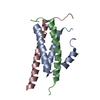

 Mass: 18.015 Da / Num. of mol.: 58 / Source method: isolated from a natural source / Formula: H2O
 Sample preparation
 Processing