 Movie
Movie Controller


+ Open data

- Basic information
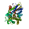
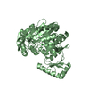
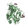
| Entry | Database: PDB / ID: 1dg6 | ||||||
|---|---|---|---|---|---|---|---|
| Title | CRYSTAL STRUCTURE OF APO2L/TRAIL | ||||||
 Components Components | APO2L/TNF-RELATED APOPOTIS INDUCING LIGAND (TRAIL) | ||||||
 Keywords Keywords | APOPTOSIS / CYTOKINE / TNF / TRIMER / ZINC-BINDING SITE | ||||||
| Function / homology |  Function and homology information Function and homology informationTRAIL binding / TRAIL signaling / TRAIL-activated apoptotic signaling pathway / Regulation by c-FLIP / CASP8 activity is inhibited / Dimerization of procaspase-8 / Caspase activation via Death Receptors in the presence of ligand / tumor necrosis factor receptor binding / positive regulation of extrinsic apoptotic signaling pathway / RIPK1-mediated regulated necrosis ...TRAIL binding / TRAIL signaling / TRAIL-activated apoptotic signaling pathway / Regulation by c-FLIP / CASP8 activity is inhibited / Dimerization of procaspase-8 / Caspase activation via Death Receptors in the presence of ligand / tumor necrosis factor receptor binding / positive regulation of extrinsic apoptotic signaling pathway / RIPK1-mediated regulated necrosis / positive regulation of release of cytochrome c from mitochondria / cytokine activity / cell-cell signaling / positive regulation of canonical NF-kappaB signal transduction / cell surface receptor signaling pathway / immune response / positive regulation of apoptotic process / signaling receptor binding / apoptotic process / signal transduction / : / extracellular exosome / extracellular region / zinc ion binding / identical protein binding / plasma membrane Similarity search - Function | ||||||
| Biological species |  Homo sapiens (human) Homo sapiens (human) | ||||||
| Method |  X-RAY DIFFRACTION / SYNCHROTRON / MOLECULAR REPLACEMENT / Resolution: 1.3 Å X-RAY DIFFRACTION / SYNCHROTRON / MOLECULAR REPLACEMENT / Resolution: 1.3 Å | ||||||
 Authors Authors | Hymowitz, S.G. / O'ConnelL, M.P. / Ultsch, M.H. / de Vos, A.M. / Kelley, R.F. | ||||||
 Citation Citation | Journal: Biochemistry / Year: 2000 Title: A unique zinc-binding site revealed by a high-resolution X-ray structure of homotrimeric Apo2L/TRAIL. Authors: Hymowitz, S.G. / O'Connell, M.P. / Ultsch, M.H. / Hurst, A. / Totpal, K. / Ashkenazi, A. / de Vos, A.M. / Kelley, R.F. | ||||||
| History |
|
- Structure visualization
| Structure viewer | Molecule: MolmilJmol/JSmol |
|---|
- Downloads & links
-Download
| PDBx/mmCIF format | 1dg6.cif.gz | 53.5 KB | Display | PDBx/mmCIF format |
|---|---|---|---|---|
| PDB format | pdb1dg6.ent.gz | 36 KB | Display | PDB format |
| PDBx/mmJSON format | 1dg6.json.gz | Tree view | PDBx/mmJSON format | |
| Others |  Other downloads Other downloads |
-Validation report
| Arichive directory | https://data.pdbj.org/pub/pdb/validation_reports/dg/1dg6ftp://data.pdbj.org/pub/pdb/validation_reports/dg/1dg6 | HTTPS FTP |
|---|
-Related structure data
| Related structure data |  1tnfS S: Starting model for refinement |
|---|---|
| Similar structure data |


-Links
 PDBj
PDBj


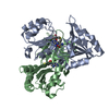
- Assembly
| Deposited unit | 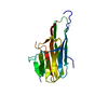
| |||||||||||||||||||||||||||||||||
|---|---|---|---|---|---|---|---|---|---|---|---|---|---|---|---|---|---|---|---|---|---|---|---|---|---|---|---|---|---|---|---|---|---|---|
| 1 | 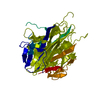
| |||||||||||||||||||||||||||||||||
| Unit cell |
| |||||||||||||||||||||||||||||||||
| Components on special symmetry positions |
|
-Components
| #1: Protein | Mass: 22106.832 Da / Num. of mol.: 1 / Fragment: TRIMERIC JELLY-ROLL DOMAIN OF APO2L / Mutation: D218A Source method: isolated from a genetically manipulated source Source: (gene. exp.) Homo sapiens (human) / Tissue: HUMAN PLACENTA / Description: HUMAN PLACENTA / Production host:  |
|---|---|
| #2: Chemical | ChemComp-ZN /   Mass: 65.409 Da / Num. of mol.: 1 / Source method: obtained synthetically / Formula: Zn Mass: 65.409 Da / Num. of mol.: 1 / Source method: obtained synthetically / Formula: Zn |
| #3: Chemical | ChemComp-CL /   Mass: 35.453 Da / Num. of mol.: 1 / Source method: obtained synthetically / Formula: Cl Mass: 35.453 Da / Num. of mol.: 1 / Source method: obtained synthetically / Formula: Cl |
| #4: Water | ChemComp-HOH /  Mass: 18.015 Da / Num. of mol.: 226 / Source method: isolated from a natural source / Formula: H2O Mass: 18.015 Da / Num. of mol.: 226 / Source method: isolated from a natural source / Formula: H2O |
-Experimental details
-Experiment
| Experiment | Method: X-RAY DIFFRACTION / Number of used crystals: 1 |
|---|
- Sample preparation
| Crystal | Density Matthews: 1.9 Å3/Da / Density % sol: 32 % | |||||||||||||||||||||||||
|---|---|---|---|---|---|---|---|---|---|---|---|---|---|---|---|---|---|---|---|---|---|---|---|---|---|---|
| Crystal grow | Temperature: 277 K / Method: vapor diffusion, sitting drop / pH: 7.5 Details: WELL SOLUTION 32% MPD; DROP 1.2MG/ML APO2L, 20MM TRIS HCL PH 7.5, 1% MPD, VAPOR DIFFUSION, SITTING DROP, temperature 277K | |||||||||||||||||||||||||
| Crystal grow | *PLUS Temperature: 4 ℃ | |||||||||||||||||||||||||
| Components of the solutions | *PLUS
|
-Data collection
| Diffraction | Mean temperature: 93 K |
|---|---|
| Diffraction source | Source: SYNCHROTRON / Site: APS  / Beamline: 19-ID / Wavelength: 1.03321 / Beamline: 19-ID / Wavelength: 1.03321 |
| Detector | Type: CUSTOM-MADE / Detector: CCD / Date: Apr 23, 1999 |
| Radiation | Protocol: SINGLE WAVELENGTH / Monochromatic (M) / Laue (L): M / Scattering type: x-ray |
| Radiation wavelength | Wavelength: 1.03321 Å / Relative weight: 1 |
| Reflection | Resolution: 1.3→20 Å / Num. all: 41901 / Num. obs: 41840 / % possible obs: 100 % / Observed criterion σ(F): 0 / Observed criterion σ(I): -3 / Redundancy: 12.1 % / Biso Wilson estimate: 12.5 Å2 / Rsym value: 6.4 / Net I/σ(I): 12.4 |
| Reflection shell | Resolution: 1.3→1.35 Å / Redundancy: 4.5 % / Rsym value: 34 / % possible all: 100 |
| Reflection | *PLUS Rmerge(I) obs: 0.064 |
| Reflection shell | *PLUS Rmerge(I) obs: 0.34 |
- Processing
| Software |
| |||||||||||||||||||||||||||||||||
|---|---|---|---|---|---|---|---|---|---|---|---|---|---|---|---|---|---|---|---|---|---|---|---|---|---|---|---|---|---|---|---|---|---|---|
| Refinement | Method to determine structure: MOLECULAR REPLACEMENT Starting model: truncated model of tnfa pdb code 1tnf Resolution: 1.3→20 Å / Num. parameters: 13240 / Num. restraintsaints: 16022 / Cross valid method: THROUGHOUT / σ(F): 0 / Stereochemistry target values: ENGH & HUBER Details: USED CONJUGANT GRADIENT LEAST SQUARES AND ANISOTROPIC B-FACTOR REFINEMENT
| |||||||||||||||||||||||||||||||||
| Solvent computation | Solvent model: Moews & Krestsinger, J.MOL.BIOL.91(1973)201-228 | |||||||||||||||||||||||||||||||||
| Refine analyze | Num. disordered residues: 5 / Occupancy sum hydrogen: 0 / Occupancy sum non hydrogen: 1452 | |||||||||||||||||||||||||||||||||
| Refinement step | Cycle: LAST / Resolution: 1.3→20 Å
| |||||||||||||||||||||||||||||||||
| Refine LS restraints |
| |||||||||||||||||||||||||||||||||
| Software | *PLUS Name: SHELXL-97 / Classification: refinement | |||||||||||||||||||||||||||||||||
| Refinement | *PLUS Lowest resolution: 20 Å / σ(F): 0 / % reflection Rfree: 10 % | |||||||||||||||||||||||||||||||||
| Solvent computation | *PLUS | |||||||||||||||||||||||||||||||||
| Displacement parameters | *PLUS | |||||||||||||||||||||||||||||||||
| Refine LS restraints | *PLUS
|
