 Movie
Movie Controller

[English] 日本語
 Yorodumi
- PDB-6kw6: Crystal Structure of cytidine deaminase from Streptomyces noursei -
+ Open data

- Basic information
| Entry | Database: PDB / ID: 6kw6 | ||||||
|---|---|---|---|---|---|---|---|
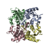
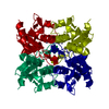
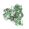
| Title | Crystal Structure of cytidine deaminase from Streptomyces noursei | ||||||
 Components Components | cytidine deaminase | ||||||
 Keywords Keywords | HYDROLASE | ||||||
| Biological species |  Streptomyces noursei (bacteria) Streptomyces noursei (bacteria) | ||||||
| Method |  X-RAY DIFFRACTION / SYNCHROTRON / MOLECULAR REPLACEMENT / molecular replacement / Resolution: 1.895 Å X-RAY DIFFRACTION / SYNCHROTRON / MOLECULAR REPLACEMENT / molecular replacement / Resolution: 1.895 Å | ||||||
 Authors Authors | Xie, T. / Liu, Z.C. / Wang, G.G. | ||||||
 Citation Citation | Journal: To Be Published Title: Crystal Structure of cytidine deaminase from Streptomyces noursei Authors: Xie, T. / Liu, Z.C. / Wang, G.G. | ||||||
| History |
|
- Structure visualization
| Structure viewer | Molecule:  MolmilJmol/JSmol MolmilJmol/JSmol |
|---|
- Downloads & links
-Download
| PDBx/mmCIF format | 6kw6.cif.gz | 60.6 KB | Display | PDBx/mmCIF format |
|---|---|---|---|---|
| PDB format | pdb6kw6.ent.gz | 42 KB | Display | PDB format |
| PDBx/mmJSON format | 6kw6.json.gz | Tree view | PDBx/mmJSON format | |
| Others |  Other downloads Other downloads |
-Validation report
| Arichive directory | https://data.pdbj.org/pub/pdb/validation_reports/kw/6kw6ftp://data.pdbj.org/pub/pdb/validation_reports/kw/6kw6 | HTTPS FTP |
|---|
-Related structure data
| Related structure data | 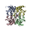 3mpzS S: Starting model for refinement |
|---|---|
| Similar structure data |


-Links
 PDBj
PDBj- Assembly
| Deposited unit | 
| |||||||||
|---|---|---|---|---|---|---|---|---|---|---|
| 1 | 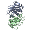
| |||||||||
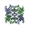
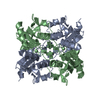
| Unit cell |
| |||||||||
| Components on special symmetry positions |
|
-Components
| #1: Protein | Mass: 13979.904 Da / Num. of mol.: 2 Source method: isolated from a genetically manipulated source Source: (gene. exp.) Streptomyces noursei (bacteria) / Production host: #2: Chemical |   Mass: 65.409 Da / Num. of mol.: 2 / Source method: obtained synthetically / Formula: Zn Mass: 65.409 Da / Num. of mol.: 2 / Source method: obtained synthetically / Formula: Zn#3: Water | ChemComp-HOH / |  Mass: 18.015 Da / Num. of mol.: 154 / Source method: isolated from a natural source / Formula: H2O Mass: 18.015 Da / Num. of mol.: 154 / Source method: isolated from a natural source / Formula: H2OHas ligand of interest | N | |
|---|
-Experimental details
-Experiment
| Experiment | Method: X-RAY DIFFRACTION / Number of used crystals: 1 |
|---|
- Sample preparation
| Crystal | Density Matthews: 2.28 Å3/Da / Density % sol: 45.97 % |
|---|---|
| Crystal grow | Temperature: 291 K / Method: vapor diffusion, hanging drop / pH: 8.5 Details: 0.2M Magnesium chloride hexahydrate, 0.1M Tris pH8.5 and 25% w/v Polyethylene glycol 3,350 |
-Data collection
| Diffraction | Mean temperature: 100 K / Serial crystal experiment: N |
|---|---|
| Diffraction source | Source: SYNCHROTRON / Site: SSRF  / Beamline: BL17U / Wavelength: 0.97916 Å / Beamline: BL17U / Wavelength: 0.97916 Å |
| Detector | Type: DECTRIS EIGER X 16M / Detector: PIXEL / Date: May 2, 2019 |
| Radiation | Protocol: SINGLE WAVELENGTH / Monochromatic (M) / Laue (L): M / Scattering type: x-ray |
| Radiation wavelength | Wavelength: 0.97916 Å / Relative weight: 1 |
| Reflection | Resolution: 1.89→29.933 Å / Num. obs: 19489 / % possible obs: 98.2 % / Redundancy: 3.3 % / Rmerge(I) obs: 0.074 / Net I/σ(I): 14.2 |
| Reflection shell | Resolution: 1.89→1.93 Å / Rmerge(I) obs: 0.476 / Mean I/σ(I) obs: 3.3 / Num. unique obs: 992 |
-Phasing
| Phasing | Method: molecular replacement |
|---|
- Processing
| Software |
| ||||||||||||||||||||||||||||||||||||||||||||||||
|---|---|---|---|---|---|---|---|---|---|---|---|---|---|---|---|---|---|---|---|---|---|---|---|---|---|---|---|---|---|---|---|---|---|---|---|---|---|---|---|---|---|---|---|---|---|---|---|---|---|
| Refinement | Method to determine structure: MOLECULAR REPLACEMENT Starting model: 3mpz Resolution: 1.895→29.933 Å / SU ML: 0.16 / Cross valid method: THROUGHOUT / σ(F): 1.36 / Phase error: 21.09
| ||||||||||||||||||||||||||||||||||||||||||||||||
| Solvent computation | Shrinkage radii: 0.9 Å / VDW probe radii: 1.11 Å | ||||||||||||||||||||||||||||||||||||||||||||||||
| Displacement parameters | Biso max: 63.22 Å2 / Biso mean: 31.2394 Å2 / Biso min: 19.66 Å2 | ||||||||||||||||||||||||||||||||||||||||||||||||
| Refinement step | Cycle: final / Resolution: 1.895→29.933 Å
| ||||||||||||||||||||||||||||||||||||||||||||||||
| Refine LS restraints |
| ||||||||||||||||||||||||||||||||||||||||||||||||
| LS refinement shell | Refine-ID: X-RAY DIFFRACTION / Rfactor Rfree error: 0
|
