 Movie
Movie Controller


+ Open data

- Basic information
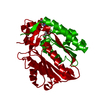
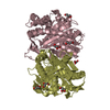
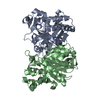
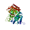
| Entry | Database: PDB / ID: 6kvs | ||||||
|---|---|---|---|---|---|---|---|
| Title | Staphylococcus aureus FabH with covalent inhibitor Oxa1 | ||||||
 Components Components | 3-oxoacyl-[acyl-carrier-protein] synthase 3 | ||||||
 Keywords Keywords | TRANSFERASE/TRANSFERASE INHIBITOR / Covalent inhibitor / Complex / ketoacyl-ACP synthase III / ANTIBIOTIC / TRANSFERASE-TRANSFERASE INHIBITOR complex | ||||||
| Function / homology |  Function and homology information Function and homology informationbeta-ketoacyl-[acyl-carrier-protein] synthase III / beta-ketoacyl-acyl-carrier-protein synthase III activity / 3-oxoacyl-[acyl-carrier-protein] synthase activity / fatty acid biosynthetic process / cytoplasm Similarity search - Function | ||||||
| Biological species |   Staphylococcus aureus (bacteria) Staphylococcus aureus (bacteria) | ||||||
| Method |  X-RAY DIFFRACTION / MOLECULAR REPLACEMENT / Resolution: 2.3 Å X-RAY DIFFRACTION / MOLECULAR REPLACEMENT / Resolution: 2.3 Å | ||||||
 Authors Authors | Yuan, Y. / Wang, J. | ||||||
 Citation Citation | Journal: To Be Published Title: Crystal Structure of S. aureus FabH, beta-ketoacyl carrier protein synthase Authors: Qiu, X. / Choudhry, A.E. | ||||||
| History |
|
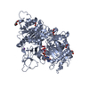
- Structure visualization
| Structure viewer | Molecule: MolmilJmol/JSmol |
|---|
- Downloads & links
-Download
| PDBx/mmCIF format | 6kvs.cif.gz | 137.1 KB | Display | PDBx/mmCIF format |
|---|---|---|---|---|
| PDB format | pdb6kvs.ent.gz | 105.2 KB | Display | PDB format |
| PDBx/mmJSON format | 6kvs.json.gz | Tree view | PDBx/mmJSON format | |
| Others |  Other downloads Other downloads |
-Validation report
| Arichive directory | https://data.pdbj.org/pub/pdb/validation_reports/kv/6kvsftp://data.pdbj.org/pub/pdb/validation_reports/kv/6kvs | HTTPS FTP |
|---|
-Related structure data
| Related structure data | 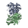 1zowS S: Starting model for refinement |
|---|---|
| Similar structure data |


-Links
 PDBj
PDBj

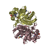
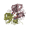
- Assembly
| Deposited unit | 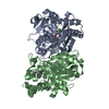
| ||||||||
|---|---|---|---|---|---|---|---|---|---|
| 1 |
| ||||||||
| Unit cell |
|
-Components
| #1: Protein | Mass: 33916.398 Da / Num. of mol.: 2 Source method: isolated from a genetically manipulated source Source: (gene. exp.) Staphylococcus aureus (strain Mu3 / ATCC 700698) (bacteria)Strain: Mu3 / ATCC 700698 / Gene: fabH, SAHV_0978 / Production host: References: UniProt: A7X0K2, beta-ketoacyl-[acyl-carrier-protein] synthase III #2: Chemical | 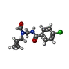  Mass: 266.723 Da / Num. of mol.: 2 / Source method: obtained synthetically / Formula: C13H15ClN2O2 Mass: 266.723 Da / Num. of mol.: 2 / Source method: obtained synthetically / Formula: C13H15ClN2O2#3: Water | ChemComp-HOH / |  Mass: 18.015 Da / Num. of mol.: 261 / Source method: isolated from a natural source / Formula: H2O Mass: 18.015 Da / Num. of mol.: 261 / Source method: isolated from a natural source / Formula: H2OHas ligand of interest | Y | Has protein modification | Y | |
|---|
-Experimental details
-Experiment
| Experiment | Method: X-RAY DIFFRACTION / Number of used crystals: 1 |
|---|
- Sample preparation
| Crystal | Density Matthews: 2.4 Å3/Da / Density % sol: 48.75 % |
|---|---|
| Crystal grow | Temperature: 291.15 K / Method: vapor diffusion, hanging drop / Details: PEG 4000 sodium citrate ammoinium acetate / PH range: 5.4-5.8 |
-Data collection
| Diffraction | Mean temperature: 100 K / Serial crystal experiment: N |
|---|---|
| Diffraction source | Source: SEALED TUBE / Type: OXFORD DIFFRACTION NOVA / Wavelength: 1.54056 Å |
| Detector | Type: RIGAKU HyPic-6000HE / Detector: PIXEL / Date: Dec 19, 2018 |
| Radiation | Protocol: SINGLE WAVELENGTH / Monochromatic (M) / Laue (L): M / Scattering type: x-ray |
| Radiation wavelength | Wavelength: 1.54056 Å / Relative weight: 1 |
| Reflection | Resolution: 2.3→22.4 Å / Num. obs: 29342 / % possible obs: 98.6 % / Redundancy: 4.6 % / Rmerge(I) obs: 0.067 / Net I/σ(I): 14.39 |
| Reflection shell | Resolution: 2.3→2.38 Å / Rmerge(I) obs: 0.344 / Num. unique obs: 2936 |
- Processing
| Software |
| ||||||||||||||||||||||||
|---|---|---|---|---|---|---|---|---|---|---|---|---|---|---|---|---|---|---|---|---|---|---|---|---|---|
| Refinement | Method to determine structure: MOLECULAR REPLACEMENT Starting model: 1ZOW Resolution: 2.3→22.14 Å / Cor.coef. Fo:Fc: 0.929 / Cor.coef. Fo:Fc free: 0.868 / SU B: 0.003 / SU ML: 0 / Cross valid method: THROUGHOUT / σ(F): 0 / ESU R: 0.2 / ESU R Free: 0.264 / Details: U VALUES : REFINED INDIVIDUALLY
| ||||||||||||||||||||||||
| Solvent computation | Ion probe radii: 0.8 Å / Shrinkage radii: 0.8 Å / VDW probe radii: 1.2 Å | ||||||||||||||||||||||||
| Displacement parameters | Biso max: 79.18 Å2 / Biso mean: 25.789 Å2 / Biso min: 9.87 Å2
| ||||||||||||||||||||||||
| Refinement step | Cycle: final / Resolution: 2.3→22.14 Å
| ||||||||||||||||||||||||
| LS refinement shell | Resolution: 2.3→2.359 Å / Rfactor Rfree error: 0 / Total num. of bins used: 20
|
