 Movie
Movie Controller


+ Open data

- Basic information
| Entry | Database: PDB / ID: 6kvc | ||||||
|---|---|---|---|---|---|---|---|
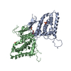
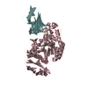
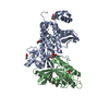
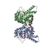
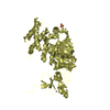
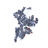
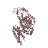
| Title | MoeE5 in complex with UDP-glucose and NAD | ||||||
 Components Components | MoeE5 | ||||||
 Keywords Keywords | BIOSYNTHETIC PROTEIN / substrate / complex / antibiotic / epimerase / galacturonic acid | ||||||
| Function / homology |  Function and homology information Function and homology informationUDP-galactose 4-epimerase, domain 1 / UDP-galactose 4-epimerase; domain 1 / NAD-dependent epimerase/dehydratase / NAD dependent epimerase/dehydratase family / NAD(P)-binding Rossmann-like Domain / NAD(P)-binding domain superfamily / Alpha-Beta Complex / Rossmann fold / 3-Layer(aba) Sandwich / Alpha Beta Similarity search - Domain/homology | ||||||
| Biological species |  Streptomyces viridosporus ATCC 14672 (bacteria) Streptomyces viridosporus ATCC 14672 (bacteria) | ||||||
| Method |  X-RAY DIFFRACTION / SYNCHROTRON / MOLECULAR REPLACEMENT / Resolution: 1.66 Å X-RAY DIFFRACTION / SYNCHROTRON / MOLECULAR REPLACEMENT / Resolution: 1.66 Å | ||||||
 Authors Authors | Ko, T.-P. / Liu, W. / Sun, H. / Liu, W. / Chen, C.-C. / Guo, R.-T. | ||||||
 Citation Citation | Journal: Biochem.Biophys.Res.Commun. / Year: 2020 Title: Structure of an antibiotic-synthesizing UDP-glucuronate 4-epimerase MoeE5 in complex with substrate. Authors: Sun, H. / Ko, T.P. / Liu, W. / Liu, W. / Zheng, Y. / Chen, C.C. / Guo, R.T. | ||||||
| History |
|
- Structure visualization
| Structure viewer | Molecule: MolmilJmol/JSmol |
|---|
- Downloads & links
-Download
| PDBx/mmCIF format | 6kvc.cif.gz | 104.3 KB | Display | PDBx/mmCIF format |
|---|---|---|---|---|
| PDB format | pdb6kvc.ent.gz | 61.8 KB | Display | PDB format |
| PDBx/mmJSON format | 6kvc.json.gz | Tree view | PDBx/mmJSON format | |
| Others |  Other downloads Other downloads |
-Validation report
| Summary document | 6kvc_validation.pdf.gz | 1 MB | Display | wwPDB validaton report |
|---|---|---|---|---|
| Full document | 6kvc_full_validation.pdf.gz | 1.1 MB | Display | |
| Data in XML | 6kvc_validation.xml.gz | 18.6 KB | Display | |
| Data in CIF | 6kvc_validation.cif.gz | 28.6 KB | Display | |
| Arichive directory | https://data.pdbj.org/pub/pdb/validation_reports/kv/6kvcftp://data.pdbj.org/pub/pdb/validation_reports/kv/6kvc | HTTPS FTP |
-Related structure data
| Related structure data | 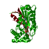 6kv9C 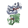 4zrnS C: citing same article ( S: Starting model for refinement |
|---|---|
| Similar structure data |


-Links
 PDBj
PDBj
- Assembly
| Deposited unit | 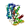
| ||||||||||||
|---|---|---|---|---|---|---|---|---|---|---|---|---|---|
| 1 | 
| ||||||||||||
| Unit cell |
| ||||||||||||
| Components on special symmetry positions |
|
-Components
| #1: Protein | Mass: 36544.141 Da / Num. of mol.: 1 Source method: isolated from a genetically manipulated source Source: (gene. exp.) Streptomyces viridosporus ATCC 14672 (bacteria)Gene: moeE5 Production host: References: UniProt: A0A003 |
|---|---|
| #2: Chemical | ChemComp-NAD /   Mass: 663.425 Da / Num. of mol.: 1 / Source method: obtained synthetically / Formula: C21H27N7O14P2 / Feature type: SUBJECT OF INVESTIGATION / Comment: NAD*YM Mass: 663.425 Da / Num. of mol.: 1 / Source method: obtained synthetically / Formula: C21H27N7O14P2 / Feature type: SUBJECT OF INVESTIGATION / Comment: NAD*YM |
| #3: Chemical | ChemComp-UPG /   Mass: 566.302 Da / Num. of mol.: 1 / Source method: obtained synthetically / Formula: C15H24N2O17P2 / Feature type: SUBJECT OF INVESTIGATION Mass: 566.302 Da / Num. of mol.: 1 / Source method: obtained synthetically / Formula: C15H24N2O17P2 / Feature type: SUBJECT OF INVESTIGATION |
| #4: Water | ChemComp-HOH /  Mass: 18.015 Da / Num. of mol.: 394 / Source method: isolated from a natural source / Formula: H2O Mass: 18.015 Da / Num. of mol.: 394 / Source method: isolated from a natural source / Formula: H2O |
| Has ligand of interest | Y |
-Experimental details
-Experiment
| Experiment | Method: X-RAY DIFFRACTION / Number of used crystals: 1 |
|---|
- Sample preparation
| Crystal | Density Matthews: 2.36 Å3/Da / Density % sol: 47.9 % |
|---|---|
| Crystal grow | Temperature: 298 K / Method: vapor diffusion, sitting drop / pH: 6.5 / Details: Na-citrate, Bis-Tris propane, PEG 3350 |
-Data collection
| Diffraction | Mean temperature: 100 K / Serial crystal experiment: N |
|---|---|
| Diffraction source | Source: SYNCHROTRON / Site: NSRRC  / Beamline: BL15A1 / Wavelength: 1 Å / Beamline: BL15A1 / Wavelength: 1 Å |
| Detector | Type: RAYONIX MX300HE / Detector: CCD / Date: Apr 27, 2016 |
| Radiation | Protocol: SINGLE WAVELENGTH / Monochromatic (M) / Laue (L): M / Scattering type: x-ray |
| Radiation wavelength | Wavelength: 1 Å / Relative weight: 1 |
| Reflection | Resolution: 1.66→25 Å / Num. obs: 42601 / % possible obs: 99.8 % / Redundancy: 10.9 % / Biso Wilson estimate: 22.06 Å2 / Rmerge(I) obs: 0.078 / Net I/σ(I): 24.5 |
| Reflection shell | Resolution: 1.66→1.72 Å / Redundancy: 7.4 % / Rmerge(I) obs: 0.563 / Mean I/σ(I) obs: 3.7 / Num. unique obs: 4165 / % possible all: 99.8 |
- Processing
| Software |
| |||||||||||||||||||||||||||||||||||||||||||||||||||||||||||||||||||||||||||||||||||||||||||||||||||||||||
|---|---|---|---|---|---|---|---|---|---|---|---|---|---|---|---|---|---|---|---|---|---|---|---|---|---|---|---|---|---|---|---|---|---|---|---|---|---|---|---|---|---|---|---|---|---|---|---|---|---|---|---|---|---|---|---|---|---|---|---|---|---|---|---|---|---|---|---|---|---|---|---|---|---|---|---|---|---|---|---|---|---|---|---|---|---|---|---|---|---|---|---|---|---|---|---|---|---|---|---|---|---|---|---|---|---|---|
| Refinement | Method to determine structure: MOLECULAR REPLACEMENT Starting model: PDB 4ZRN Resolution: 1.66→24.75 Å / SU ML: 0.1382 / Cross valid method: THROUGHOUT / σ(F): 1.34 / Phase error: 18.5802
| |||||||||||||||||||||||||||||||||||||||||||||||||||||||||||||||||||||||||||||||||||||||||||||||||||||||||
| Solvent computation | Shrinkage radii: 0.9 Å / VDW probe radii: 1.11 Å | |||||||||||||||||||||||||||||||||||||||||||||||||||||||||||||||||||||||||||||||||||||||||||||||||||||||||
| Displacement parameters | Biso mean: 26.46 Å2 | |||||||||||||||||||||||||||||||||||||||||||||||||||||||||||||||||||||||||||||||||||||||||||||||||||||||||
| Refinement step | Cycle: LAST / Resolution: 1.66→24.75 Å
| |||||||||||||||||||||||||||||||||||||||||||||||||||||||||||||||||||||||||||||||||||||||||||||||||||||||||
| Refine LS restraints |
| |||||||||||||||||||||||||||||||||||||||||||||||||||||||||||||||||||||||||||||||||||||||||||||||||||||||||
| LS refinement shell |
|
