 Movie
Movie Controller

[English] 日本語
 Yorodumi
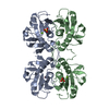
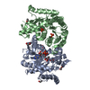
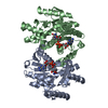
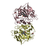
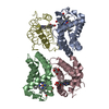
- PDB-6kp5: crystal structure of Xanthine-guanine phosphoribosyltransferase (... -
+ Open data

- Basic information
| Entry | Database: PDB / ID: 6kp5 | ||||||
|---|---|---|---|---|---|---|---|
| Title | crystal structure of Xanthine-guanine phosphoribosyltransferase (XGPRT) from Yersinia pestis in P21212 space group with sulphate ions in the active site | ||||||
 Components Components | Xanthine phosphoribosyltransferase | ||||||
 Keywords Keywords | TRANSFERASE / xanthine-guanine phosphoribosyltransferase / Yersinia pestis | ||||||
| Function / homology |  Function and homology information Function and homology informationxanthine phosphoribosyltransferase / XMP salvage / xanthine phosphoribosyltransferase activity / guanine phosphoribosyltransferase activity / hypoxanthine phosphoribosyltransferase activity / GMP salvage / IMP salvage / purine ribonucleoside salvage / Transferases; Glycosyltransferases; Pentosyltransferases / magnesium ion binding ...xanthine phosphoribosyltransferase / XMP salvage / xanthine phosphoribosyltransferase activity / guanine phosphoribosyltransferase activity / hypoxanthine phosphoribosyltransferase activity / GMP salvage / IMP salvage / purine ribonucleoside salvage / Transferases; Glycosyltransferases; Pentosyltransferases / magnesium ion binding / plasma membrane / cytosol Similarity search - Function | ||||||
| Biological species |   Yersinia pestis (bacteria) Yersinia pestis (bacteria) | ||||||
| Method |  X-RAY DIFFRACTION / SYNCHROTRON / MOLECULAR REPLACEMENT / Resolution: 1.1 Å X-RAY DIFFRACTION / SYNCHROTRON / MOLECULAR REPLACEMENT / Resolution: 1.1 Å | ||||||
 Authors Authors | Lankipalli, S. / Ramagopal, U.A. | ||||||
 Citation Citation | Journal: To be published Title: crystal structure of Xanthine-guanine phosphoribosyltransferase (XGPRT) from Yersinia pestis in P21212 space group Authors: Lankipalli, S. / Ramagopal, U.A. | ||||||
| History |
|
- Structure visualization
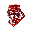
| Structure viewer | Molecule: MolmilJmol/JSmol |
|---|
- Downloads & links
-Download
| PDBx/mmCIF format | 6kp5.cif.gz | 133.9 KB | Display | PDBx/mmCIF format |
|---|---|---|---|---|
| PDB format | pdb6kp5.ent.gz | 103.3 KB | Display | PDB format |
| PDBx/mmJSON format | 6kp5.json.gz | Tree view | PDBx/mmJSON format | |
| Others |  Other downloads Other downloads |
-Validation report
| Arichive directory | https://data.pdbj.org/pub/pdb/validation_reports/kp/6kp5ftp://data.pdbj.org/pub/pdb/validation_reports/kp/6kp5 | HTTPS FTP |
|---|
-Related structure data
| Related structure data |  5xtkS S: Starting model for refinement |
|---|---|
| Similar structure data |


-Links
 PDBj
PDBj
- Assembly
| Deposited unit | 
| ||||||||||||||||||
|---|---|---|---|---|---|---|---|---|---|---|---|---|---|---|---|---|---|---|---|
| 1 | 
| ||||||||||||||||||
| Unit cell |
| ||||||||||||||||||
| Components on special symmetry positions |
|
-Components
| #1: Protein | Mass: 17072.594 Da / Num. of mol.: 2 Source method: isolated from a genetically manipulated source Source: (gene. exp.) Yersinia pestis (bacteria) / Gene: gpt, YPO3225, y0963, YP_0708 / Plasmid: pNIC28-Bsa4 / Production host: References: UniProt: Q8ZC05, xanthine phosphoribosyltransferase #2: Chemical | ChemComp-SO4 /   Mass: 96.063 Da / Num. of mol.: 4 / Source method: obtained synthetically / Formula: SO4 / Feature type: SUBJECT OF INVESTIGATION Mass: 96.063 Da / Num. of mol.: 4 / Source method: obtained synthetically / Formula: SO4 / Feature type: SUBJECT OF INVESTIGATION#3: Water | ChemComp-HOH / |  Mass: 18.015 Da / Num. of mol.: 228 / Source method: isolated from a natural source / Formula: H2O Mass: 18.015 Da / Num. of mol.: 228 / Source method: isolated from a natural source / Formula: H2OHas ligand of interest | Y | |
|---|
-Experimental details
-Experiment
| Experiment | Method: X-RAY DIFFRACTION / Number of used crystals: 1 |
|---|
- Sample preparation
| Crystal | Density Matthews: 2.09 Å3/Da / Density % sol: 41.09 % / Mosaicity: 0.23 ° |
|---|---|
| Crystal grow | Temperature: 298 K / Method: vapor diffusion, sitting drop / pH: 8.5 Details: 0.2M Lithium sulphate, 0.1M Tris:HCl pH 8.5, 1.26M Ammonium Sulfate |
-Data collection
| Diffraction | Mean temperature: 100 K / Serial crystal experiment: N | ||||||||||||||||||||||||
|---|---|---|---|---|---|---|---|---|---|---|---|---|---|---|---|---|---|---|---|---|---|---|---|---|---|
| Diffraction source | Source: SYNCHROTRON / Site: ELETTRA  / Beamline: 11.2C / Wavelength: 0.9762 Å / Beamline: 11.2C / Wavelength: 0.9762 Å | ||||||||||||||||||||||||
| Detector | Type: DECTRIS PILATUS 6M / Detector: PIXEL / Date: Dec 6, 2018 | ||||||||||||||||||||||||
| Radiation | Protocol: SINGLE WAVELENGTH / Monochromatic (M) / Laue (L): M / Scattering type: x-ray | ||||||||||||||||||||||||
| Radiation wavelength | Wavelength: 0.9762 Å / Relative weight: 1 | ||||||||||||||||||||||||
| Reflection | Resolution: 1.1→47.32 Å / Num. obs: 116357 / % possible obs: 99.9 % / Redundancy: 11.3 % / CC1/2: 0.998 / Rmerge(I) obs: 0.112 / Rpim(I) all: 0.035 / Rrim(I) all: 0.117 / Net I/σ(I): 10.4 | ||||||||||||||||||||||||
| Reflection shell | Diffraction-ID: 1
|
- Processing
| Software |
| |||||||||||||||||||||||||||||||||||||||||||||||||||||||||||||||||
|---|---|---|---|---|---|---|---|---|---|---|---|---|---|---|---|---|---|---|---|---|---|---|---|---|---|---|---|---|---|---|---|---|---|---|---|---|---|---|---|---|---|---|---|---|---|---|---|---|---|---|---|---|---|---|---|---|---|---|---|---|---|---|---|---|---|---|
| Refinement | Method to determine structure: MOLECULAR REPLACEMENT Starting model: 5XTK Resolution: 1.1→47.315 Å / Cor.coef. Fo:Fc: 0.97 / Cor.coef. Fo:Fc free: 0.963 / SU B: 1.346 / SU ML: 0.029 / Cross valid method: THROUGHOUT / σ(F): 0 / ESU R: 0.031 / ESU R Free: 0.031 Details: HYDROGENS HAVE BEEN ADDED IN THE RIDING POSITIONS U VALUES : REFINED INDIVIDUALLY
| |||||||||||||||||||||||||||||||||||||||||||||||||||||||||||||||||
| Solvent computation | Ion probe radii: 0.8 Å / Shrinkage radii: 0.8 Å / VDW probe radii: 1.2 Å | |||||||||||||||||||||||||||||||||||||||||||||||||||||||||||||||||
| Displacement parameters | Biso max: 66.01 Å2 / Biso mean: 12.589 Å2 / Biso min: 5.62 Å2
| |||||||||||||||||||||||||||||||||||||||||||||||||||||||||||||||||
| Refinement step | Cycle: final / Resolution: 1.1→47.315 Å
| |||||||||||||||||||||||||||||||||||||||||||||||||||||||||||||||||
| Refine LS restraints |
| |||||||||||||||||||||||||||||||||||||||||||||||||||||||||||||||||
| LS refinement shell | Resolution: 1.1→1.129 Å / Rfactor Rfree error: 0 / Total num. of bins used: 20
|
