 Movie
Movie Controller

[English] 日本語
 Yorodumi
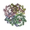
- PDB-1gv0: Structural Basis for Thermophilic Protein Stability: Structures o... -
+ Open data

- Basic information
| Entry | Database: PDB / ID: 1gv0 | |||||||||
|---|---|---|---|---|---|---|---|---|---|---|
| Title | Structural Basis for Thermophilic Protein Stability: Structures of Thermophilic and Mesophilic Malate Dehydrogenases | |||||||||
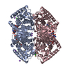
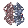
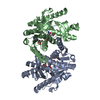
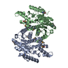
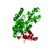
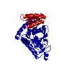
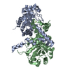
 Components Components | MALATE DEHYDROGENASE | |||||||||
 Keywords Keywords | OXIDOREDUCTASE / DEHYDROGENASE / TRICARBOXYLIC ACID / NAD | |||||||||
| Function / homology |  Function and homology information Function and homology information(S)-malate dehydrogenase (NAD+, oxaloacetate-forming) / L-malate dehydrogenase (NAD+) activity / L-lactate dehydrogenase (NAD+) activity / lactate metabolic process / tricarboxylic acid cycle Similarity search - Function | |||||||||
| Biological species |  CHLOROBIUM TEPIDUM (bacteria) CHLOROBIUM TEPIDUM (bacteria) | |||||||||
| Method |  X-RAY DIFFRACTION / SYNCHROTRON / MOLECULAR REPLACEMENT / Resolution: 2.5 Å X-RAY DIFFRACTION / SYNCHROTRON / MOLECULAR REPLACEMENT / Resolution: 2.5 Å | |||||||||
 Authors Authors | Dalhus, B. / Sarinen, M. / Sauer, U.H. / Eklund, P. / Johansson, K. / Karlsson, A. / Ramaswamy, S. / Bjork, A. / Synstad, B. / Naterstad, K. ...Dalhus, B. / Sarinen, M. / Sauer, U.H. / Eklund, P. / Johansson, K. / Karlsson, A. / Ramaswamy, S. / Bjork, A. / Synstad, B. / Naterstad, K. / Sirevag, R. / Eklund, H. | |||||||||
 Citation Citation | Journal: J.Mol.Biol. / Year: 2002 Title: Structural Basis for Thermophilic Protein Stability: Structures of Thermophilic and Mesophilic Malate Dehydrogenases Authors: Dalhus, B. / Saarinen, M. / Sauer, U.H. / Eklund, P. / Johansson, K. / Karlsson, A. / Ramaswamy, S. / Bjork, A. / Synstad, B. / Naterstad, K. / Sirevag, R. / Eklund, H. | |||||||||
| History |
|
- Structure visualization
| Structure viewer | Molecule: MolmilJmol/JSmol |
|---|
- Downloads & links
-Download
| PDBx/mmCIF format | 1gv0.cif.gz | 129 KB | Display | PDBx/mmCIF format |
|---|---|---|---|---|
| PDB format | pdb1gv0.ent.gz | 100.5 KB | Display | PDB format |
| PDBx/mmJSON format | 1gv0.json.gz | Tree view | PDBx/mmJSON format | |
| Others |  Other downloads Other downloads |
-Validation report
| Arichive directory | https://data.pdbj.org/pub/pdb/validation_reports/gv/1gv0ftp://data.pdbj.org/pub/pdb/validation_reports/gv/1gv0 | HTTPS FTP |
|---|
-Related structure data
| Related structure data | 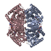 1guyC 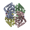 1guzSC 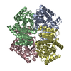 1gv1C C: citing same article ( S: Starting model for refinement |
|---|---|
| Similar structure data |

-Links
 PDBj
PDBj


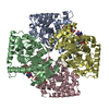
- Assembly
| Deposited unit | 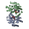
| ||||||||
|---|---|---|---|---|---|---|---|---|---|
| 1 | 
| ||||||||
| Unit cell |
|
-Components
| #1: Protein | Mass: 33144.230 Da / Num. of mol.: 2 Source method: isolated from a genetically manipulated source Source: (gene. exp.) CHLOROBIUM TEPIDUM (bacteria) / Production host: References: UniProt: P80039, (S)-malate dehydrogenase (NAD+, oxaloacetate-forming) #2: Chemical |   Mass: 663.425 Da / Num. of mol.: 2 / Source method: obtained synthetically / Formula: C21H27N7O14P2 / Comment: NAD*YM Mass: 663.425 Da / Num. of mol.: 2 / Source method: obtained synthetically / Formula: C21H27N7O14P2 / Comment: NAD*YM#3: Water | ChemComp-HOH / |  Mass: 18.015 Da / Num. of mol.: 233 / Source method: isolated from a natural source / Formula: H2O Mass: 18.015 Da / Num. of mol.: 233 / Source method: isolated from a natural source / Formula: H2O |
|---|
-Experimental details
-Experiment
| Experiment | Method: X-RAY DIFFRACTION / Number of used crystals: 1 |
|---|
- Sample preparation
| Crystal | Density Matthews: 3.1 Å3/Da / Density % sol: 60 % | ||||||||||||||||||||||||||||||
|---|---|---|---|---|---|---|---|---|---|---|---|---|---|---|---|---|---|---|---|---|---|---|---|---|---|---|---|---|---|---|---|
| Crystal grow | pH: 7.5 Details: 20MG/ML,50MMTRIS-HCL,PH7.4,55%MPD, 100MMHEPES,PH7.5., pH 7.50 | ||||||||||||||||||||||||||||||
| Crystal grow | *PLUS Temperature: 15 ℃ / pH: 7.4 / Method: vapor diffusion, hanging drop | ||||||||||||||||||||||||||||||
| Components of the solutions | *PLUS
|
-Data collection
| Diffraction | Mean temperature: 100 K |
|---|---|
| Diffraction source | Source: SYNCHROTRON / Site: MAX II  / Beamline: I711 / Wavelength: 0.9831 / Beamline: I711 / Wavelength: 0.9831 |
| Detector | Type: MARRESEARCH / Detector: IMAGE PLATE |
| Radiation | Protocol: SINGLE WAVELENGTH / Monochromatic (M) / Laue (L): M / Scattering type: x-ray |
| Radiation wavelength | Wavelength: 0.9831 Å / Relative weight: 1 |
| Reflection | Resolution: 2.5→40.8 Å / Num. obs: 28459 / % possible obs: 100 % / Observed criterion σ(I): 0 / Redundancy: 2.5 % / Biso Wilson estimate: 14.5 Å2 / Rmerge(I) obs: 0.07 |
| Reflection shell | Resolution: 2.5→2.64 Å / Rmerge(I) obs: 0.254 / % possible all: 100 |
| Reflection | *PLUS % possible obs: 100 % / Num. measured all: 117908 / Rmerge(I) obs: 0.07 |
| Reflection shell | *PLUS % possible obs: 100 % |
- Processing
| Software |
| ||||||||||||||||||||||||||||||||||||||||||||||||||||||||||||||||||||||||||||||||
|---|---|---|---|---|---|---|---|---|---|---|---|---|---|---|---|---|---|---|---|---|---|---|---|---|---|---|---|---|---|---|---|---|---|---|---|---|---|---|---|---|---|---|---|---|---|---|---|---|---|---|---|---|---|---|---|---|---|---|---|---|---|---|---|---|---|---|---|---|---|---|---|---|---|---|---|---|---|---|---|---|---|
| Refinement | Method to determine structure: MOLECULAR REPLACEMENT Starting model: MDH WITH PDB-CODE 1GUZ Resolution: 2.5→40.9 Å / Rfactor Rfree error: 0.005 / Data cutoff high absF: 3775346.2 / Isotropic thermal model: RESTRAINED / Cross valid method: THROUGHOUT / σ(F): 0
| ||||||||||||||||||||||||||||||||||||||||||||||||||||||||||||||||||||||||||||||||
| Solvent computation | Solvent model: FLAT MODEL / Bsol: 50.952 Å2 / ksol: 0.371756 e/Å3 | ||||||||||||||||||||||||||||||||||||||||||||||||||||||||||||||||||||||||||||||||
| Displacement parameters | Biso mean: 32.8 Å2
| ||||||||||||||||||||||||||||||||||||||||||||||||||||||||||||||||||||||||||||||||
| Refine analyze |
| ||||||||||||||||||||||||||||||||||||||||||||||||||||||||||||||||||||||||||||||||
| Refinement step | Cycle: LAST / Resolution: 2.5→40.9 Å
| ||||||||||||||||||||||||||||||||||||||||||||||||||||||||||||||||||||||||||||||||
| Refine LS restraints |
| ||||||||||||||||||||||||||||||||||||||||||||||||||||||||||||||||||||||||||||||||
| LS refinement shell | Resolution: 2.5→2.66 Å / Rfactor Rfree error: 0.018 / Total num. of bins used: 6
| ||||||||||||||||||||||||||||||||||||||||||||||||||||||||||||||||||||||||||||||||
| Xplor file |
| ||||||||||||||||||||||||||||||||||||||||||||||||||||||||||||||||||||||||||||||||
| Software | *PLUS Name: CNS / Version: 1 / Classification: refinement | ||||||||||||||||||||||||||||||||||||||||||||||||||||||||||||||||||||||||||||||||
| Refinement | *PLUS | ||||||||||||||||||||||||||||||||||||||||||||||||||||||||||||||||||||||||||||||||
| Solvent computation | *PLUS | ||||||||||||||||||||||||||||||||||||||||||||||||||||||||||||||||||||||||||||||||
| Displacement parameters | *PLUS Biso mean: 31.4 Å2 | ||||||||||||||||||||||||||||||||||||||||||||||||||||||||||||||||||||||||||||||||
| Refine LS restraints | *PLUS
|
