 Movie
Movie Controller


+ Open data

- Basic information
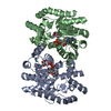
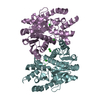
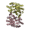
| Entry | Database: PDB / ID: 1nul | ||||||
|---|---|---|---|---|---|---|---|
| Title | XPRTASE FROM E. COLI | ||||||
 Components Components | XANTHINE-GUANINE PHOSPHORIBOSYLTRANSFERASE | ||||||
 Keywords Keywords | PHOSPHORIBOSYLTRANSFERASE / TRANSFERASE / PURINE SALVAGE ENZYME | ||||||
| Function / homology |  Function and homology information Function and homology informationxanthine phosphoribosyltransferase / XMP salvage / xanthine phosphoribosyltransferase activity / guanine phosphoribosyltransferase activity / hypoxanthine phosphoribosyltransferase activity / GMP salvage / IMP salvage / purine ribonucleoside salvage / guanosine tetraphosphate binding / Transferases; Glycosyltransferases; Pentosyltransferases ...xanthine phosphoribosyltransferase / XMP salvage / xanthine phosphoribosyltransferase activity / guanine phosphoribosyltransferase activity / hypoxanthine phosphoribosyltransferase activity / GMP salvage / IMP salvage / purine ribonucleoside salvage / guanosine tetraphosphate binding / Transferases; Glycosyltransferases; Pentosyltransferases / protein homotetramerization / magnesium ion binding / protein-containing complex / identical protein binding / plasma membrane / cytosol Similarity search - Function | ||||||
| Biological species |  | ||||||
| Method |  X-RAY DIFFRACTION / MIR / Resolution: 1.8 Å X-RAY DIFFRACTION / MIR / Resolution: 1.8 Å | ||||||
 Authors Authors | Vos, S. / De Jersey, J. / Martin, J.L. | ||||||
 Citation Citation | Journal: Biochemistry / Year: 1997 Title: Crystal structure of Escherichia coli xanthine phosphoribosyltransferase. Authors: Vos, S. / de Jersey, J. / Martin, J.L. #1: Journal: J.Struct.Biol. / Year: 1996Title: Crystallization and Preliminary X-Ray Crystallographic Studies of Escherichia Coli Xanthine Phosphoribosyltransferase Authors: Vos, S. / De Jersey, J. / Martin, J.L. | ||||||
| History |
|
- Structure visualization
| Structure viewer | Molecule: MolmilJmol/JSmol |
|---|
- Downloads & links
-Download
| PDBx/mmCIF format | 1nul.cif.gz | 71.6 KB | Display | PDBx/mmCIF format |
|---|---|---|---|---|
| PDB format | pdb1nul.ent.gz | 52.7 KB | Display | PDB format |
| PDBx/mmJSON format | 1nul.json.gz | Tree view | PDBx/mmJSON format | |
| Others |  Other downloads Other downloads |
-Validation report
| Arichive directory | https://data.pdbj.org/pub/pdb/validation_reports/nu/1nulftp://data.pdbj.org/pub/pdb/validation_reports/nu/1nul | HTTPS FTP |
|---|
-Related structure data
| Similar structure data |
|---|


-Links
 PDBj
PDBj
- Assembly
| Deposited unit | 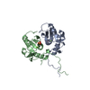
| ||||||||||||
|---|---|---|---|---|---|---|---|---|---|---|---|---|---|
| 1 | 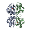
| ||||||||||||
| Unit cell |
| ||||||||||||
| Components on special symmetry positions |
| ||||||||||||
| Noncrystallographic symmetry (NCS) | NCS oper: (Code: given Matrix: (-0.0901, -0.9906, 0.1028), Vector: |
-Components
| #1: Protein | Mass: 16991.568 Da / Num. of mol.: 2 / Source method: isolated from a natural source / Source: (natural) References: UniProt: P0A9M5, xanthine phosphoribosyltransferase #2: Chemical |   Mass: 96.063 Da / Num. of mol.: 2 / Source method: obtained synthetically / Formula: SO4 Mass: 96.063 Da / Num. of mol.: 2 / Source method: obtained synthetically / Formula: SO4#3: Chemical |   Mass: 24.305 Da / Num. of mol.: 2 / Source method: obtained synthetically / Formula: Mg Mass: 24.305 Da / Num. of mol.: 2 / Source method: obtained synthetically / Formula: Mg#4: Water | ChemComp-HOH / |  Mass: 18.015 Da / Num. of mol.: 212 / Source method: isolated from a natural source / Formula: H2O Mass: 18.015 Da / Num. of mol.: 212 / Source method: isolated from a natural source / Formula: H2O |
|---|
-Experimental details
-Experiment
| Experiment | Method: X-RAY DIFFRACTION / Number of used crystals: 1 |
|---|
- Sample preparation
| Crystal | Density Matthews: 2.15 Å3/Da / Density % sol: 43 % | ||||||||||||||||||||||||
|---|---|---|---|---|---|---|---|---|---|---|---|---|---|---|---|---|---|---|---|---|---|---|---|---|---|
| Crystal grow | pH: 8 Details: XPRT WAS CRYSTALLISED FROM 18 - 23% PEG 4000, 0.1 M LI2SO4 IN 0.1 M TRIS-HCL, PH 8., pH 8.0 | ||||||||||||||||||||||||
| Crystal grow | *PLUS Method: vapor diffusion, hanging drop | ||||||||||||||||||||||||
| Components of the solutions | *PLUS
|
-Data collection
| Diffraction | Mean temperature: 289 K |
|---|---|
| Diffraction source | Source: ROTATING ANODE / Type: RIGAKU RUH2R / Wavelength: 1.5418 |
| Detector | Type: RIGAKU / Detector: IMAGE PLATE / Date: May 1, 1995 / Details: MIRRORS |
| Radiation | Monochromator: NI FILTER / Monochromatic (M) / Laue (L): M / Scattering type: x-ray |
| Radiation wavelength | Wavelength: 1.5418 Å / Relative weight: 1 |
| Reflection | Resolution: 1.8→53.2 Å / Num. obs: 25622 / % possible obs: 92 % / Observed criterion σ(I): 1 / Redundancy: 3 % / Biso Wilson estimate: 19.5 Å2 / Rmerge(I) obs: 0.06 / Net I/σ(I): 14.8 |
| Reflection shell | Resolution: 1.8→2 Å / Redundancy: 2.4 % / Rmerge(I) obs: 0.263 / Mean I/σ(I) obs: 3.2 / % possible all: 84 |
| Reflection | *PLUS Num. measured all: 77524 / Rmerge(I) obs: 0.06 |
| Reflection shell | *PLUS % possible obs: 81.9 % |
- Processing
| Software |
| ||||||||||||||||||||||||||||||||||||||||||||||||||||||||||||||||||||||||||||||||
|---|---|---|---|---|---|---|---|---|---|---|---|---|---|---|---|---|---|---|---|---|---|---|---|---|---|---|---|---|---|---|---|---|---|---|---|---|---|---|---|---|---|---|---|---|---|---|---|---|---|---|---|---|---|---|---|---|---|---|---|---|---|---|---|---|---|---|---|---|---|---|---|---|---|---|---|---|---|---|---|---|---|
| Refinement | Method to determine structure: MIR / Resolution: 1.8→8 Å / Rfactor Rfree error: 0.005 / Data cutoff high absF: 1644 / Data cutoff low absF: 16.6 / Cross valid method: THROUGHOUT / σ(F): 2
| ||||||||||||||||||||||||||||||||||||||||||||||||||||||||||||||||||||||||||||||||
| Displacement parameters | Biso mean: 25.5 Å2
| ||||||||||||||||||||||||||||||||||||||||||||||||||||||||||||||||||||||||||||||||
| Refine analyze | Luzzati coordinate error obs: 0.24 Å / Luzzati d res low obs: 5 Å / Luzzati sigma a obs: 0.29 Å | ||||||||||||||||||||||||||||||||||||||||||||||||||||||||||||||||||||||||||||||||
| Refinement step | Cycle: LAST / Resolution: 1.8→8 Å
| ||||||||||||||||||||||||||||||||||||||||||||||||||||||||||||||||||||||||||||||||
| Refine LS restraints |
| ||||||||||||||||||||||||||||||||||||||||||||||||||||||||||||||||||||||||||||||||
| LS refinement shell | Resolution: 1.8→1.9 Å / Rfactor Rfree error: 0.019
| ||||||||||||||||||||||||||||||||||||||||||||||||||||||||||||||||||||||||||||||||
| Xplor file |
| ||||||||||||||||||||||||||||||||||||||||||||||||||||||||||||||||||||||||||||||||
| Software | *PLUS Name: X-PLOR / Version: 3.1 / Classification: refinement | ||||||||||||||||||||||||||||||||||||||||||||||||||||||||||||||||||||||||||||||||
| Refine LS restraints | *PLUS
|
