 Movie
Movie Controller


+ Open data

- Basic information
| Entry | Database: PDB / ID: 6ko6 | ||||||
|---|---|---|---|---|---|---|---|
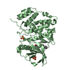
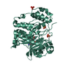
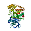
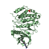
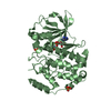
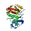
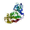
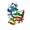
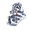
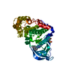
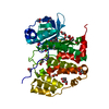
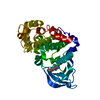
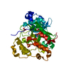
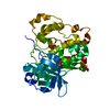
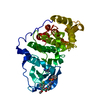
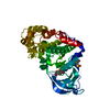
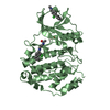
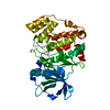
| Title | Crystal structure of AMPPNP bound Cka1 from C. neoformans | ||||||
 Components Components | CMGC/CK2 protein kinase | ||||||
 Keywords Keywords | TRANSFERASE / kinase | ||||||
| Function / homology |  Function and homology information Function and homology informationregulation of transcription by RNA polymerase III / regulation of transcription by RNA polymerase I / protein kinase CK2 complex / non-specific serine/threonine protein kinase / regulation of cell cycle / protein serine kinase activity / protein serine/threonine kinase activity / DNA damage response / ATP binding / metal ion binding ...regulation of transcription by RNA polymerase III / regulation of transcription by RNA polymerase I / protein kinase CK2 complex / non-specific serine/threonine protein kinase / regulation of cell cycle / protein serine kinase activity / protein serine/threonine kinase activity / DNA damage response / ATP binding / metal ion binding / nucleus / cytosol Similarity search - Function | ||||||
| Biological species |  Cryptococcus neoformans var. grubii serotype A (fungus) Cryptococcus neoformans var. grubii serotype A (fungus) | ||||||
| Method |  X-RAY DIFFRACTION / SYNCHROTRON / MOLECULAR REPLACEMENT / Resolution: 2.4 Å X-RAY DIFFRACTION / SYNCHROTRON / MOLECULAR REPLACEMENT / Resolution: 2.4 Å | ||||||
 Authors Authors | Cho, H.S. / Yoo, Y. | ||||||
 Citation Citation | Journal: Sci Rep / Year: 2019 Title: Structural analysis of fungal pathogenicity-related casein kinase alpha subunit, Cka1, in the human fungal pathogen Cryptococcus neoformans. Authors: Ong, B.X. / Yoo, Y. / Han, M.G. / Park, J.B. / Choi, M.K. / Choi, Y. / Shin, J.S. / Bahn, Y.S. / Cho, H.S. #1: Journal: J.Mol.Biol. / Year: 2007Title: Evolved to be active: sulfate ions define substrate recognition sites of CK2alpha and emphasise its exceptional role within the CMGC family of eukaryotic protein kinases. Authors: Niefind, K. / Yde, C.W. / Ermakova, I. / Issinger, O.G. | ||||||
| History |
|
- Structure visualization
| Structure viewer | Molecule: MolmilJmol/JSmol |
|---|
- Downloads & links
-Download
| PDBx/mmCIF format | 6ko6.cif.gz | 86.6 KB | Display | PDBx/mmCIF format |
|---|---|---|---|---|
| PDB format | pdb6ko6.ent.gz | 62.5 KB | Display | PDB format |
| PDBx/mmJSON format | 6ko6.json.gz | Tree view | PDBx/mmJSON format | |
| Others |  Other downloads Other downloads |
-Validation report
| Arichive directory | https://data.pdbj.org/pub/pdb/validation_reports/ko/6ko6ftp://data.pdbj.org/pub/pdb/validation_reports/ko/6ko6 | HTTPS FTP |
|---|
-Related structure data
| Related structure data |  6k3lC  1lp4S S: Starting model for refinement C: citing same article ( |
|---|---|
| Similar structure data |


-Links
 PDBj
PDBj


- Assembly
| Deposited unit | 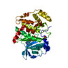
| ||||||||||||
|---|---|---|---|---|---|---|---|---|---|---|---|---|---|
| 1 |
| ||||||||||||
| Unit cell |
|
-Components
| #1: Protein | Mass: 39580.965 Da / Num. of mol.: 1 Source method: isolated from a genetically manipulated source Source: (gene. exp.) Cryptococcus neoformans var. grubii serotype A (strain H99 / ATCC 208821 / CBS 10515 / FGSC 9487) (fungus)Strain: H99 / ATCC 208821 / CBS 10515 / FGSC 9487 / Gene: CNAG_05694 Production host:  References: UniProt: J9VNH4, UniProt: A0A226BA59*PLUS | ||||
|---|---|---|---|---|---|
| #2: Chemical | ChemComp-ANP /   Mass: 506.196 Da / Num. of mol.: 1 / Source method: obtained synthetically / Formula: C10H17N6O12P3 / Feature type: SUBJECT OF INVESTIGATION / Comment: AMP-PNP, energy-carrying molecule analogue*YM Mass: 506.196 Da / Num. of mol.: 1 / Source method: obtained synthetically / Formula: C10H17N6O12P3 / Feature type: SUBJECT OF INVESTIGATION / Comment: AMP-PNP, energy-carrying molecule analogue*YM | ||||
| #3: Chemical | ChemComp-MG /   Mass: 24.305 Da / Num. of mol.: 1 / Source method: obtained synthetically / Formula: Mg Mass: 24.305 Da / Num. of mol.: 1 / Source method: obtained synthetically / Formula: Mg | ||||
| #4: Chemical |   Mass: 96.063 Da / Num. of mol.: 2 / Source method: obtained synthetically / Formula: SO4 Mass: 96.063 Da / Num. of mol.: 2 / Source method: obtained synthetically / Formula: SO4#5: Water | ChemComp-HOH / |  Mass: 18.015 Da / Num. of mol.: 25 / Source method: isolated from a natural source / Formula: H2O Mass: 18.015 Da / Num. of mol.: 25 / Source method: isolated from a natural source / Formula: H2OHas ligand of interest | Y | |
-Experimental details
-Experiment
| Experiment | Method: X-RAY DIFFRACTION / Number of used crystals: 1 |
|---|
- Sample preparation
| Crystal | Density Matthews: 2.51 Å3/Da / Density % sol: 51.07 % |
|---|---|
| Crystal grow | Temperature: 290 K / Method: vapor diffusion, sitting drop / pH: 8.5 Details: 0.2M lithium sulfate, 0.1M Tris pH 8.5, 30%(w/v) PEG 4000, 30%(w/v) dextran sulfate sodium salt, 5mM AMPPNP, 5mM MgCl2 |
-Data collection
| Diffraction | Mean temperature: 100 K / Serial crystal experiment: N |
|---|---|
| Diffraction source | Source: SYNCHROTRON / Site: Photon Factory  / Beamline: BL-1A / Wavelength: 0.97 Å / Beamline: BL-1A / Wavelength: 0.97 Å |
| Detector | Type: DECTRIS EIGER X 4M / Detector: PIXEL / Date: May 27, 2019 |
| Radiation | Protocol: SINGLE WAVELENGTH / Monochromatic (M) / Laue (L): M / Scattering type: x-ray |
| Radiation wavelength | Wavelength: 0.97 Å / Relative weight: 1 |
| Reflection | Resolution: 2.4→46.67 Å / Num. obs: 15949 / % possible obs: 99.5 % / Redundancy: 2 % / CC1/2: 0.998 / Rmerge(I) obs: 0.0417 / Net I/σ(I): 10.6 |
| Reflection shell | Resolution: 2.4→2.486 Å / Rmerge(I) obs: 0.286 / Mean I/σ(I) obs: 2.35 / Num. unique obs: 1554 / CC1/2: 0.929 |
- Processing
| Software |
| ||||||||||||||||
|---|---|---|---|---|---|---|---|---|---|---|---|---|---|---|---|---|---|
| Refinement | Method to determine structure: MOLECULAR REPLACEMENT Starting model: 1LP4 Resolution: 2.4→46.67 Å / Cross valid method: THROUGHOUT
| ||||||||||||||||
| Refinement step | Cycle: LAST / Resolution: 2.4→46.67 Å
| ||||||||||||||||
| LS refinement shell | Resolution: 2.4→2.486 Å /
|
