[English] 日本語
 Yorodumi
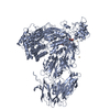
- PDB-6kez: Crystal structure of GAPDH/CP12/PRK complex from Arabidopsis thaliana -
+ Open data
- Basic information
| Entry | Database: PDB / ID: 6kez | ||||||
|---|---|---|---|---|---|---|---|
| Title | Crystal structure of GAPDH/CP12/PRK complex from Arabidopsis thaliana | ||||||
 Components Components |
| ||||||
 Keywords Keywords | OXIDOREDUCTASE/TRANSFERASE/PROTEIN BINDING / glyceraldehyde-2-phosphate dehydrogenase / phosphoribulokinase / CP12 chloroplast / OXIDOREDUCTASE-TRANSFERASE-PROTEIN BINDING complex | ||||||
| Function / homology |  Function and homology information Function and homology informationphosphoribulokinase / phosphoribulokinase activity / negative regulation of reductive pentose-phosphate cycle / cellular response to anoxia / stromule / glyceraldehyde-3-phosphate dehydrogenase (NADP+) (phosphorylating) / salicylic acid binding / glyceraldehyde-3-phosphate dehydrogenase (NADP+) (phosphorylating) activity / chloroplast membrane / response to sucrose ...phosphoribulokinase / phosphoribulokinase activity / negative regulation of reductive pentose-phosphate cycle / cellular response to anoxia / stromule / glyceraldehyde-3-phosphate dehydrogenase (NADP+) (phosphorylating) / salicylic acid binding / glyceraldehyde-3-phosphate dehydrogenase (NADP+) (phosphorylating) activity / chloroplast membrane / response to sucrose / thylakoid / apoplast / reductive pentose-phosphate cycle / glyceraldehyde-3-phosphate dehydrogenase (NAD+) (phosphorylating) activity / chloroplast envelope / chloroplast stroma / cellular response to cold / chloroplast thylakoid membrane / nickel cation binding / response to light stimulus / response to cold / chloroplast / glucose metabolic process / NAD binding / disordered domain specific binding / NADP binding / cellular response to heat / protein-containing complex assembly / protein-macromolecule adaptor activity / protein homotetramerization / copper ion binding / mRNA binding / protein-containing complex binding / enzyme binding / protein homodimerization activity / protein-containing complex / ATP binding / identical protein binding / nucleus / cytoplasm / cytosol Similarity search - Function | ||||||
| Biological species |  | ||||||
| Method |  X-RAY DIFFRACTION / X-RAY DIFFRACTION /  SYNCHROTRON / SYNCHROTRON /  MOLECULAR REPLACEMENT / Resolution: 3.5 Å MOLECULAR REPLACEMENT / Resolution: 3.5 Å | ||||||
 Authors Authors | Yu, A. / Xie, Y. / Li, M. | ||||||
| Funding support |  China, 1items China, 1items
| ||||||
 Citation Citation |  Journal: Plant Cell / Year: 2020 Journal: Plant Cell / Year: 2020Title: Photosynthetic Phosphoribulokinase Structures: Enzymatic Mechanisms and the Redox Regulation of the Calvin-Benson-Bassham Cycle. Authors: Yu, A. / Xie, Y. / Pan, X. / Zhang, H. / Cao, P. / Su, X. / Chang, W. / Li, M. | ||||||
| History |
|
- Structure visualization
| Structure viewer | Molecule:  Molmil Molmil Jmol/JSmol Jmol/JSmol |
|---|
- Downloads & links
- Download
| PDBx/mmCIF format |  6kez.cif.gz 6kez.cif.gz | 798 KB | Display |  PDBx/mmCIF format PDBx/mmCIF format |
|---|---|---|---|---|
| PDB format |  pdb6kez.ent.gz pdb6kez.ent.gz | 661.7 KB | Display |  PDB format PDB format |
| PDBx/mmJSON format |  6kez.json.gz 6kez.json.gz | Tree view |  PDBx/mmJSON format PDBx/mmJSON format | |
| Others |  Other downloads Other downloads |
-Validation report
| Arichive directory |  https://data.pdbj.org/pub/pdb/validation_reports/ke/6kez https://data.pdbj.org/pub/pdb/validation_reports/ke/6kez ftp://data.pdbj.org/pub/pdb/validation_reports/ke/6kez ftp://data.pdbj.org/pub/pdb/validation_reports/ke/6kez | HTTPS FTP |
|---|
-Related structure data
| Related structure data | 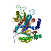 6kevC  6kewSC  6kexC  3rvdS S: Starting model for refinement C: citing same article ( |
|---|---|
| Similar structure data |
- Links
Links
- Assembly
| Deposited unit | 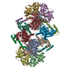
| ||||||||||||
|---|---|---|---|---|---|---|---|---|---|---|---|---|---|
| 1 | 
| ||||||||||||
| 2 | 
| ||||||||||||
| Unit cell |
|
- Components
| #1: Protein | Mass: 36592.750 Da / Num. of mol.: 8 Source method: isolated from a genetically manipulated source Source: (gene. exp.)   References: UniProt: P25856, glyceraldehyde-3-phosphate dehydrogenase (NADP+) (phosphorylating) #2: Protein | Mass: 39514.785 Da / Num. of mol.: 4 Source method: isolated from a genetically manipulated source Source: (gene. exp.)   #3: Protein | Mass: 8542.076 Da / Num. of mol.: 4 Source method: isolated from a genetically manipulated source Source: (gene. exp.)   #4: Chemical | ChemComp-NAD / #5: Water | ChemComp-HOH / | Has ligand of interest | Y | Has protein modification | Y | |
|---|
-Experimental details
-Experiment
| Experiment | Method:  X-RAY DIFFRACTION / Number of used crystals: 1 X-RAY DIFFRACTION / Number of used crystals: 1 |
|---|
- Sample preparation
| Crystal | Density Matthews: 3 Å3/Da / Density % sol: 58.97 % |
|---|---|
| Crystal grow | Temperature: 291.15 K / Method: vapor diffusion, sitting drop Details: 100mM HEPES, pH7.5, 100mM KCl, 15% polyethylene glycol 6000 |
-Data collection
| Diffraction | Mean temperature: 100 K / Serial crystal experiment: N |
|---|---|
| Diffraction source | Source:  SYNCHROTRON / Site: SYNCHROTRON / Site:  SSRF SSRF  / Beamline: BL17U / Wavelength: 0.98 Å / Beamline: BL17U / Wavelength: 0.98 Å |
| Detector | Type: DECTRIS EIGER X 16M / Detector: PIXEL / Date: Mar 21, 2018 |
| Radiation | Protocol: SINGLE WAVELENGTH / Monochromatic (M) / Laue (L): M / Scattering type: x-ray |
| Radiation wavelength | Wavelength: 0.98 Å / Relative weight: 1 |
| Reflection | Resolution: 3.5→50 Å / Num. obs: 68615 / % possible obs: 97.8 % / Redundancy: 3.3 % / Biso Wilson estimate: 55.99606247 Å2 / Rmerge(I) obs: 0.123 / Net I/σ(I): 7.56 |
| Reflection shell | Resolution: 3.5→3.56 Å / Rmerge(I) obs: 0.554 / Num. unique obs: 3452 |
- Processing
| Software |
| ||||||||||||||||
|---|---|---|---|---|---|---|---|---|---|---|---|---|---|---|---|---|---|
| Refinement | Method to determine structure:  MOLECULAR REPLACEMENT MOLECULAR REPLACEMENTStarting model: 3RVD, 6KEW Resolution: 3.5→50 Å / Cross valid method: FREE R-VALUE
| ||||||||||||||||
| Refinement step | Cycle: LAST / Resolution: 3.5→50 Å
| ||||||||||||||||
| LS refinement shell | Resolution: 3.497→3.622 Å /
|
 Movie
Movie Controller




 PDBj
PDBj


