 Movie
Movie Controller

[English] 日本語
 Yorodumi
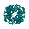
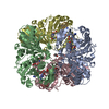
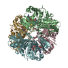
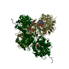
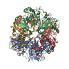
- PDB-3rvd: Crystal structure of the binary complex, obtained by soaking, of ... -
+ Open data

- Basic information
| Entry | Database: PDB / ID: 3rvd | ||||||
|---|---|---|---|---|---|---|---|
| Title | Crystal structure of the binary complex, obtained by soaking, of photosyntetic a4 glyceraldehyde 3-phosphate dehydrogenase (gapdh) with cp12-2, both from arabidopsis thaliana. | ||||||
 Components Components |
| ||||||
 Keywords Keywords | OXIDOREDUCTASE/PROTEIN BINDING / Rossmann fold / Calvin cycle / binary complex / chloroplast / OXIDOREDUCTASE-PROTEIN BINDING complex | ||||||
| Function / homology |  Function and homology information Function and homology informationnegative regulation of reductive pentose-phosphate cycle / cellular response to anoxia / stromule / glyceraldehyde-3-phosphate dehydrogenase (NADP+) (phosphorylating) / salicylic acid binding / glyceraldehyde-3-phosphate dehydrogenase (NADP+) (phosphorylating) activity / chloroplast membrane / response to sucrose / apoplast / reductive pentose-phosphate cycle ...negative regulation of reductive pentose-phosphate cycle / cellular response to anoxia / stromule / glyceraldehyde-3-phosphate dehydrogenase (NADP+) (phosphorylating) / salicylic acid binding / glyceraldehyde-3-phosphate dehydrogenase (NADP+) (phosphorylating) activity / chloroplast membrane / response to sucrose / apoplast / reductive pentose-phosphate cycle / glyceraldehyde-3-phosphate dehydrogenase (NAD+) (phosphorylating) activity / chloroplast envelope / chloroplast stroma / cellular response to cold / chloroplast thylakoid membrane / nickel cation binding / response to light stimulus / response to cold / chloroplast / glucose metabolic process / NAD binding / disordered domain specific binding / NADP binding / cellular response to heat / protein-containing complex assembly / protein homotetramerization / protein-macromolecule adaptor activity / copper ion binding / mRNA binding / protein-containing complex binding / enzyme binding / protein homodimerization activity / protein-containing complex / identical protein binding / cytosol Similarity search - Function | ||||||
| Biological species |  | ||||||
| Method |  X-RAY DIFFRACTION / SYNCHROTRON / MOLECULAR REPLACEMENT / Resolution: 2.7 Å X-RAY DIFFRACTION / SYNCHROTRON / MOLECULAR REPLACEMENT / Resolution: 2.7 Å | ||||||
 Authors Authors | Fermani, S. / Thumiger, A. / Falini, G. / Marri, L. / Sparla, F. / Trost, P. | ||||||
 Citation Citation | Journal: J.Biol.Chem. / Year: 2012 Title: Conformational Selection and Folding-upon-binding of Intrinsically Disordered Protein CP12 Regulate Photosynthetic Enzymes Assembly. Authors: Fermani, S. / Trivelli, X. / Sparla, F. / Thumiger, A. / Calvaresi, M. / Marri, L. / Falini, G. / Zerbetto, F. / Trost, P. #1: Journal: Acta Crystallogr.,Sect.F / Year: 2010Title: Structure of photosynthetic glyceraldehyde-3-phosphate dehydrogenase (isoform a4) from arabidopsis thaliana in complex with nad. Authors: Fermani, S. / Sparla, F. / Marri, L. / Tumigher, A. / Pupillo, P. / Falini, G. / Trost, P. | ||||||
| History |
|
- Structure visualization
| Structure viewer | Molecule: MolmilJmol/JSmol |
|---|
- Downloads & links
-Download
| PDBx/mmCIF format | 3rvd.cif.gz | 675.2 KB | Display | PDBx/mmCIF format |
|---|---|---|---|---|
| PDB format | pdb3rvd.ent.gz | 558 KB | Display | PDB format |
| PDBx/mmJSON format | 3rvd.json.gz | Tree view | PDBx/mmJSON format | |
| Others |  Other downloads Other downloads |
-Validation report
| Arichive directory | https://data.pdbj.org/pub/pdb/validation_reports/rv/3rvdftp://data.pdbj.org/pub/pdb/validation_reports/rv/3rvd | HTTPS FTP |
|---|
-Related structure data
| Related structure data |  2lj9C  3qv1C  3k2bS S: Starting model for refinement C: citing same article ( |
|---|---|
| Similar structure data |

-Links
 PDBj
PDBj
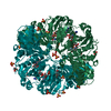
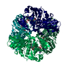
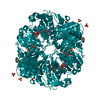
- Assembly
| Deposited unit | 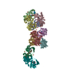
| |||||||||
|---|---|---|---|---|---|---|---|---|---|---|
| 1 | 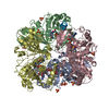
| |||||||||
| 2 | 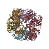
| |||||||||
| 3 | 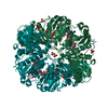
| |||||||||
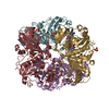
| Unit cell |
| |||||||||
| Components on special symmetry positions |
|
-Components
| #1: Protein | Mass: 36391.570 Da / Num. of mol.: 10 Source method: isolated from a genetically manipulated source Source: (gene. exp.)  References: UniProt: P25856, glyceraldehyde-3-phosphate dehydrogenase (NADP+) (phosphorylating) #2: Protein | Mass: 8754.368 Da / Num. of mol.: 6 / Fragment: UNP residues 54-131 Source method: isolated from a genetically manipulated source Source: (gene. exp.) #3: Chemical | ChemComp-NAD /   Mass: 663.425 Da / Num. of mol.: 10 / Source method: obtained synthetically / Formula: C21H27N7O14P2 / Comment: NAD*YM Mass: 663.425 Da / Num. of mol.: 10 / Source method: obtained synthetically / Formula: C21H27N7O14P2 / Comment: NAD*YM#4: Chemical | ChemComp-SO4 /   Mass: 96.063 Da / Num. of mol.: 28 / Source method: obtained synthetically / Formula: SO4 Mass: 96.063 Da / Num. of mol.: 28 / Source method: obtained synthetically / Formula: SO4#5: Water | ChemComp-HOH / |  Mass: 18.015 Da / Num. of mol.: 556 / Source method: isolated from a natural source / Formula: H2O Mass: 18.015 Da / Num. of mol.: 556 / Source method: isolated from a natural source / Formula: H2OHas protein modification | Y | |
|---|
-Experimental details
-Experiment
| Experiment | Method: X-RAY DIFFRACTION / Number of used crystals: 1 |
|---|
- Sample preparation
| Crystal | Density Matthews: 2.96 Å3/Da / Density % sol: 58.49 % |
|---|---|
| Crystal grow | Temperature: 293 K / Method: vapor diffusion, sitting drop Details: 2.4 M ammonium sulphate, 0.1 M sodium citrate, 1 mM NAD, 3.125 mg/ml CP12 for soaking experiments, VAPOR DIFFUSION, SITTING DROP, temperature 293K |
-Data collection
| Diffraction | Mean temperature: 100 K |
|---|---|
| Diffraction source | Source: SYNCHROTRON / Site: ESRF  / Beamline: ID14-1 / Wavelength: 0.934 Å / Beamline: ID14-1 / Wavelength: 0.934 Å |
| Detector | Type: ADSC QUANTUM 210 / Detector: CCD / Date: Sep 9, 2006 |
| Radiation | Protocol: SINGLE WAVELENGTH / Monochromatic (M) / Laue (L): M / Scattering type: x-ray |
| Radiation wavelength | Wavelength: 0.934 Å / Relative weight: 1 |
| Reflection | Resolution: 2.7→95.3 Å / Num. all: 119322 / Num. obs: 118509 / % possible obs: 96.6 % / Observed criterion σ(F): 3 / Observed criterion σ(I): -3 / Redundancy: 11.3 % / Biso Wilson estimate: 27.5 Å2 / Rmerge(I) obs: 0.098 / Rsym value: 0.098 / Net I/σ(I): 16.3 |
| Reflection shell | Resolution: 2.7→2.8 Å / Redundancy: 7.9 % / Rmerge(I) obs: 0.587 / Mean I/σ(I) obs: 2.1 / Rsym value: 0.587 / % possible all: 84 |
- Processing
| Software |
| ||||||||||||||||||||||||||||||||||||||||||||||||||||||||||||||||||||||||||||||||
|---|---|---|---|---|---|---|---|---|---|---|---|---|---|---|---|---|---|---|---|---|---|---|---|---|---|---|---|---|---|---|---|---|---|---|---|---|---|---|---|---|---|---|---|---|---|---|---|---|---|---|---|---|---|---|---|---|---|---|---|---|---|---|---|---|---|---|---|---|---|---|---|---|---|---|---|---|---|---|---|---|---|
| Refinement | Method to determine structure: MOLECULAR REPLACEMENT Starting model: PDB ENTRY 3K2B Resolution: 2.7→94.61 Å / Rfactor Rfree error: 0.003 / Data cutoff high absF: 3188319.93 / Data cutoff low absF: 0 / Isotropic thermal model: RESTRAINED / Cross valid method: THROUGHOUT / σ(F): 0 / Stereochemistry target values: Engh & Huber / Details: BULK SOLVENT MODEL USED
| ||||||||||||||||||||||||||||||||||||||||||||||||||||||||||||||||||||||||||||||||
| Solvent computation | Solvent model: FLAT MODEL / Bsol: 58.8944 Å2 / ksol: 0.36 e/Å3 | ||||||||||||||||||||||||||||||||||||||||||||||||||||||||||||||||||||||||||||||||
| Displacement parameters | Biso mean: 63.5 Å2
| ||||||||||||||||||||||||||||||||||||||||||||||||||||||||||||||||||||||||||||||||
| Refine analyze |
| ||||||||||||||||||||||||||||||||||||||||||||||||||||||||||||||||||||||||||||||||
| Refinement step | Cycle: LAST / Resolution: 2.7→94.61 Å
| ||||||||||||||||||||||||||||||||||||||||||||||||||||||||||||||||||||||||||||||||
| Refine LS restraints |
| ||||||||||||||||||||||||||||||||||||||||||||||||||||||||||||||||||||||||||||||||
| Refine LS restraints NCS | NCS model details: NONE | ||||||||||||||||||||||||||||||||||||||||||||||||||||||||||||||||||||||||||||||||
| LS refinement shell | Resolution: 2.7→2.87 Å / Rfactor Rfree error: 0.01 / Total num. of bins used: 6
| ||||||||||||||||||||||||||||||||||||||||||||||||||||||||||||||||||||||||||||||||
| Xplor file |
|
