 Movie
Movie Controller

[English] 日本語
 Yorodumi
- PDB-6kct: Crystal structure of plasmodium lysyl-tRNA synthetase in complex ... -
+ Open data

- Basic information
| Entry | Database: PDB / ID: 6kct | ||||||
|---|---|---|---|---|---|---|---|
| Title | Crystal structure of plasmodium lysyl-tRNA synthetase in complex with a cladosporin derivative 5 | ||||||
 Components Components | Lysine--tRNA ligase | ||||||
 Keywords Keywords | LIGASE / Inhibitor / Complex | ||||||
| Function / homology |  Function and homology information Function and homology informationlysine-tRNA ligase / lysine-tRNA ligase activity / lysyl-tRNA aminoacylation / tRNA binding / ATP binding / cytosol Similarity search - Function | ||||||
| Biological species |  | ||||||
| Method |  X-RAY DIFFRACTION / SYNCHROTRON / MOLECULAR REPLACEMENT / Resolution: 3.25 Å X-RAY DIFFRACTION / SYNCHROTRON / MOLECULAR REPLACEMENT / Resolution: 3.25 Å | ||||||
 Authors Authors | Zhou, J. / Fang, P. | ||||||
| Funding support |  China, 1items China, 1items
| ||||||
 Citation Citation | Journal: Acs Chem.Biol. / Year: 2020 Title: Atomic Resolution Analyses of Isocoumarin Derivatives for Inhibition of Lysyl-tRNA Synthetase. Authors: Zhou, J. / Zheng, L. / Hei, Z. / Li, W. / Wang, J. / Yu, B. / Fang, P. | ||||||
| History |
|
- Structure visualization
| Structure viewer | Molecule: MolmilJmol/JSmol |
|---|
- Downloads & links
-Download
| PDBx/mmCIF format | 6kct.cif.gz | 767.8 KB | Display | PDBx/mmCIF format |
|---|---|---|---|---|
| PDB format | pdb6kct.ent.gz | 635.1 KB | Display | PDB format |
| PDBx/mmJSON format | 6kct.json.gz | Tree view | PDBx/mmJSON format | |
| Others |  Other downloads Other downloads |
-Validation report
| Arichive directory | https://data.pdbj.org/pub/pdb/validation_reports/kc/6kctftp://data.pdbj.org/pub/pdb/validation_reports/kc/6kct | HTTPS FTP |
|---|
-Related structure data
| Related structure data |  6ka6C  6kabC  6kbfC  6kcnC  4ycvS S: Starting model for refinement C: citing same article ( |
|---|---|
| Similar structure data |


-Links
 PDBj
PDBj

- Assembly
| Deposited unit | 
| ||||||||
|---|---|---|---|---|---|---|---|---|---|
| 1 | 
| ||||||||
| 2 | 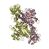
| ||||||||
| Unit cell |
|
-Components
| #1: Protein | Mass: 59781.133 Da / Num. of mol.: 4 Source method: isolated from a genetically manipulated source Source: (gene. exp.) Strain: isolate NF54 / Gene: PFNF54_04763 / Production host:  #2: Chemical | ChemComp-D5O /   Mass: 274.312 Da / Num. of mol.: 4 / Source method: obtained synthetically / Formula: C16H18O4 / Feature type: SUBJECT OF INVESTIGATION Mass: 274.312 Da / Num. of mol.: 4 / Source method: obtained synthetically / Formula: C16H18O4 / Feature type: SUBJECT OF INVESTIGATION#3: Chemical | ChemComp-LYS /   Type: L-peptide linking / Mass: 147.195 Da / Num. of mol.: 4 / Source method: isolated from a natural source / Formula: C6H15N2O2 Type: L-peptide linking / Mass: 147.195 Da / Num. of mol.: 4 / Source method: isolated from a natural source / Formula: C6H15N2O2#4: Water | ChemComp-HOH / |  Mass: 18.015 Da / Num. of mol.: 80 / Source method: isolated from a natural source / Formula: H2O Mass: 18.015 Da / Num. of mol.: 80 / Source method: isolated from a natural source / Formula: H2OHas ligand of interest | Y | |
|---|
-Experimental details
-Experiment
| Experiment | Method: X-RAY DIFFRACTION / Number of used crystals: 1 |
|---|
- Sample preparation
| Crystal | Density Matthews: 2.47 Å3/Da / Density % sol: 50.12 % |
|---|---|
| Crystal grow | Temperature: 291 K / Method: vapor diffusion, sitting drop / Details: tri-sodium citrate, PEG 3350 |
-Data collection
| Diffraction | Mean temperature: 100 K / Serial crystal experiment: N | |||||||||||||||||||||||||||||||||||||||||||||||||||||||||||||||||||||||||||||||||||||||||||||||||||
|---|---|---|---|---|---|---|---|---|---|---|---|---|---|---|---|---|---|---|---|---|---|---|---|---|---|---|---|---|---|---|---|---|---|---|---|---|---|---|---|---|---|---|---|---|---|---|---|---|---|---|---|---|---|---|---|---|---|---|---|---|---|---|---|---|---|---|---|---|---|---|---|---|---|---|---|---|---|---|---|---|---|---|---|---|---|---|---|---|---|---|---|---|---|---|---|---|---|---|---|---|
| Diffraction source | Source: SYNCHROTRON / Site: SSRF / Beamline: BL19U1 / Wavelength: 0.987 Å | |||||||||||||||||||||||||||||||||||||||||||||||||||||||||||||||||||||||||||||||||||||||||||||||||||
| Detector | Type: ADSC QUANTUM 315r / Detector: CCD / Date: Mar 24, 2019 | |||||||||||||||||||||||||||||||||||||||||||||||||||||||||||||||||||||||||||||||||||||||||||||||||||
| Radiation | Protocol: SINGLE WAVELENGTH / Monochromatic (M) / Laue (L): M / Scattering type: x-ray | |||||||||||||||||||||||||||||||||||||||||||||||||||||||||||||||||||||||||||||||||||||||||||||||||||
| Radiation wavelength | Wavelength: 0.987 Å / Relative weight: 1 | |||||||||||||||||||||||||||||||||||||||||||||||||||||||||||||||||||||||||||||||||||||||||||||||||||
| Reflection | Resolution: 3.25→50 Å / Num. obs: 33930 / % possible obs: 94.7 % / Redundancy: 3.6 % / Biso Wilson estimate: 58.99 Å2 / Rmerge(I) obs: 0.249 / Rpim(I) all: 0.159 / Rrim(I) all: 0.297 / Χ2: 1.04 / Net I/σ(I): 2.6 / Num. measured all: 121802 | |||||||||||||||||||||||||||||||||||||||||||||||||||||||||||||||||||||||||||||||||||||||||||||||||||
| Reflection shell | Diffraction-ID: 1
|
- Processing
| Software |
| |||||||||||||||||||||||||||||||||||||||||||||||||||||||||||||||||||||||||||||
|---|---|---|---|---|---|---|---|---|---|---|---|---|---|---|---|---|---|---|---|---|---|---|---|---|---|---|---|---|---|---|---|---|---|---|---|---|---|---|---|---|---|---|---|---|---|---|---|---|---|---|---|---|---|---|---|---|---|---|---|---|---|---|---|---|---|---|---|---|---|---|---|---|---|---|---|---|---|---|
| Refinement | Method to determine structure: MOLECULAR REPLACEMENT Starting model: 4YCV Resolution: 3.25→48.946 Å / SU ML: 0.54 / Cross valid method: THROUGHOUT / σ(F): 1.92 / Phase error: 38.53 / Stereochemistry target values: ML
| |||||||||||||||||||||||||||||||||||||||||||||||||||||||||||||||||||||||||||||
| Solvent computation | Shrinkage radii: 0.9 Å / VDW probe radii: 1.11 Å / Solvent model: FLAT BULK SOLVENT MODEL | |||||||||||||||||||||||||||||||||||||||||||||||||||||||||||||||||||||||||||||
| Displacement parameters | Biso max: 111.66 Å2 / Biso mean: 60.8876 Å2 / Biso min: 33.27 Å2 | |||||||||||||||||||||||||||||||||||||||||||||||||||||||||||||||||||||||||||||
| Refinement step | Cycle: final / Resolution: 3.25→48.946 Å
| |||||||||||||||||||||||||||||||||||||||||||||||||||||||||||||||||||||||||||||
| LS refinement shell | Refine-ID: X-RAY DIFFRACTION / Rfactor Rfree error: 0 / Total num. of bins used: 10
| |||||||||||||||||||||||||||||||||||||||||||||||||||||||||||||||||||||||||||||
| Refinement TLS params. | Method: refined / Origin x: 1.496 Å / Origin y: 13.1431 Å / Origin z: 22.7805 Å
| |||||||||||||||||||||||||||||||||||||||||||||||||||||||||||||||||||||||||||||
| Refinement TLS group |
|
