 Movie
Movie Controller

[English] 日本語
 Yorodumi
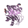
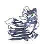
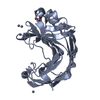
- PDB-6k98: Substrates promiscuity of xyloglucanases and endoglucanases of gl... -
+ Open data

- Basic information
| Entry | Database: PDB / ID: 6k98 | ||||||
|---|---|---|---|---|---|---|---|
| Title | Substrates promiscuity of xyloglucanases and endoglucanases of glycoside hydrolase 12 family | ||||||
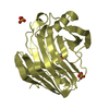
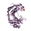
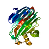
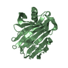
 Components Components | GH12 beta-1, 4-endoglucanase | ||||||
 Keywords Keywords | HYDROLASE / endoglucanase / glycoside hydrolase family 12 / PROTEIN FIBRIL | ||||||
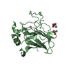
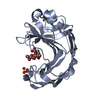
| Function / homology |  Function and homology information Function and homology information | ||||||
| Biological species |  | ||||||
| Method |  X-RAY DIFFRACTION / SYNCHROTRON / MOLECULAR REPLACEMENT / Resolution: 2.032 Å X-RAY DIFFRACTION / SYNCHROTRON / MOLECULAR REPLACEMENT / Resolution: 2.032 Å | ||||||
 Authors Authors | Hong, Y. / Tao, T. / Pengjun, S. / Jiaming, C. / Xiaoyu, W. / Chen, H. / yingguo, B. / Bin, Y. | ||||||
 Citation Citation | Journal: To Be Published Title: Substrates promiscuity of xyloglucanases and endoglucanases of glycoside hydrolase 12 family Authors: Hong, Y. / Tao, T. / Pengjun, S. / Jiaming, C. / Xiaoyu, W. / Chen, H. / yingguo, B. / Bin, Y. | ||||||
| History |
|
- Structure visualization
| Structure viewer | Molecule: MolmilJmol/JSmol |
|---|
- Downloads & links
-Download
| PDBx/mmCIF format | 6k98.cif.gz | 59.4 KB | Display | PDBx/mmCIF format |
|---|---|---|---|---|
| PDB format | pdb6k98.ent.gz | 41.6 KB | Display | PDB format |
| PDBx/mmJSON format | 6k98.json.gz | Tree view | PDBx/mmJSON format | |
| Others |  Other downloads Other downloads |
-Validation report
| Arichive directory | https://data.pdbj.org/pub/pdb/validation_reports/k9/6k98ftp://data.pdbj.org/pub/pdb/validation_reports/k9/6k98 | HTTPS FTP |
|---|
-Related structure data
| Related structure data |  6k9dC  1ks4S S: Starting model for refinement C: citing same article ( |
|---|---|
| Similar structure data |

-Links
 PDBj
PDBj
- Assembly
| Deposited unit | 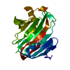
| ||||||||
|---|---|---|---|---|---|---|---|---|---|
| 1 |
| ||||||||
| Unit cell |
|
-Components
| #1: Protein | Mass: 24205.309 Da / Num. of mol.: 1 Source method: isolated from a genetically manipulated source Source: (gene. exp.) Komagataella phaffii CBS 7435 (fungus) / References: UniProt: A0A1L6CE30, cellulase |
|---|---|
| #2: Water | ChemComp-HOH /  Mass: 18.015 Da / Num. of mol.: 80 / Source method: isolated from a natural source / Formula: H2O Mass: 18.015 Da / Num. of mol.: 80 / Source method: isolated from a natural source / Formula: H2O |
| Has protein modification | Y |
-Experimental details
-Experiment
| Experiment | Method: X-RAY DIFFRACTION / Number of used crystals: 1 |
|---|
- Sample preparation
| Crystal | Density Matthews: 5.32 Å3/Da / Density % sol: 76.89 % |
|---|---|
| Crystal grow | Temperature: 298.15 K / Method: vapor diffusion, hanging drop Details: NfEG12A was concentrated to 10 mg/ml in a buffer containing 100 mM citric acid-Na2HPO4 (pH 7.2). The protein was crystallized in a mother liquid with 0.1 M citrate (pH 5.0) and 16% (w/v) PEG ...Details: NfEG12A was concentrated to 10 mg/ml in a buffer containing 100 mM citric acid-Na2HPO4 (pH 7.2). The protein was crystallized in a mother liquid with 0.1 M citrate (pH 5.0) and 16% (w/v) PEG 20000 using the hanging drop vapor diffusion method in 24-well plates. NfEG12A crystals was transferred to a reservoir solution supplemented with 25% (v/v) ethylene glycol to grown. Obtained crystals were cryoprotected with 25% (v/v) 2-methyl-2,4-pentanediol. The crystals were mounted on a nylon loop and cooled immediately in liquid nitrogen prior to data collection. |
-Data collection
| Diffraction | Mean temperature: 298 K / Serial crystal experiment: N |
|---|---|
| Diffraction source | Source: SYNCHROTRON / Site: NSRRC  / Beamline: BL15A1 / Wavelength: 1.00919 Å / Beamline: BL15A1 / Wavelength: 1.00919 Å |
| Detector | Type: DECTRIS PILATUS 6M / Detector: PIXEL / Date: Jan 4, 2019 |
| Radiation | Protocol: SINGLE WAVELENGTH / Monochromatic (M) / Laue (L): M / Scattering type: x-ray |
| Radiation wavelength | Wavelength: 1.00919 Å / Relative weight: 1 |
| Reflection | Resolution: 2.032→57.522 Å / Num. obs: 29949 / % possible obs: 87.23 % / Redundancy: 4.2 % / Net I/σ(I): 1.35 |
| Reflection shell | Resolution: 2.03255→4.519 Å / Num. unique obs: 11 |
- Processing
| Software |
| ||||||||||||||||||||||||||||||||||||||||||||||||||||||||||||||||||||||||||||||||||||
|---|---|---|---|---|---|---|---|---|---|---|---|---|---|---|---|---|---|---|---|---|---|---|---|---|---|---|---|---|---|---|---|---|---|---|---|---|---|---|---|---|---|---|---|---|---|---|---|---|---|---|---|---|---|---|---|---|---|---|---|---|---|---|---|---|---|---|---|---|---|---|---|---|---|---|---|---|---|---|---|---|---|---|---|---|---|
| Refinement | Method to determine structure: MOLECULAR REPLACEMENT Starting model: 1KS4 Resolution: 2.032→57.522 Å / SU ML: 0.24 / Cross valid method: FREE R-VALUE / σ(F): 1.35 / Phase error: 26.74
| ||||||||||||||||||||||||||||||||||||||||||||||||||||||||||||||||||||||||||||||||||||
| Solvent computation | Shrinkage radii: 0.9 Å / VDW probe radii: 1.11 Å | ||||||||||||||||||||||||||||||||||||||||||||||||||||||||||||||||||||||||||||||||||||
| Refinement step | Cycle: LAST / Resolution: 2.032→57.522 Å
| ||||||||||||||||||||||||||||||||||||||||||||||||||||||||||||||||||||||||||||||||||||
| Refine LS restraints |
| ||||||||||||||||||||||||||||||||||||||||||||||||||||||||||||||||||||||||||||||||||||
| LS refinement shell |
|
