 Movie
Movie Controller


+ Open data

- Basic information
| Entry | Database: PDB / ID: 6k1t | ||||||
|---|---|---|---|---|---|---|---|
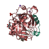
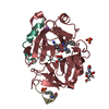
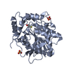
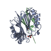
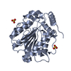
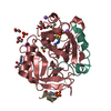
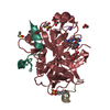
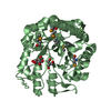
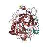
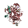
| Title | The structure of Francisella virulence factor BioJ | ||||||
 Components Components | Alpha/beta hydrolase fold family protein | ||||||
 Keywords Keywords | HYDROLASE / virulence factor BioJ | ||||||
| Function / homology | Alpha/beta hydrolase fold-3 / alpha/beta hydrolase fold / Alpha/Beta hydrolase fold / hydrolase activity / Alpha/beta hydrolase fold family protein Function and homology information Function and homology information | ||||||
| Biological species |  Francisella philomiragia subsp. philomiragia ATCC 25015 (bacteria) Francisella philomiragia subsp. philomiragia ATCC 25015 (bacteria) | ||||||
| Method |  X-RAY DIFFRACTION / SYNCHROTRON / MAD / Resolution: 1.584 Å X-RAY DIFFRACTION / SYNCHROTRON / MAD / Resolution: 1.584 Å | ||||||
 Authors Authors | Ouyang, S. / Guan, H. / Zhang, S. | ||||||
 Citation Citation | Journal: Iscience / Year: 2019 Title: Molecular Basis of BioJ, a Unique Gatekeeper in Bacterial Biotin Synthesis. Authors: Wei, W. / Guan, H. / Zhu, T. / Zhang, S. / Fan, C. / Ouyang, S. / Feng, Y. | ||||||
| History |
|
- Structure visualization
| Structure viewer | Molecule: MolmilJmol/JSmol |
|---|
- Downloads & links
-Download
| PDBx/mmCIF format | 6k1t.cif.gz | 78.2 KB | Display | PDBx/mmCIF format |
|---|---|---|---|---|
| PDB format | pdb6k1t.ent.gz | 56.6 KB | Display | PDB format |
| PDBx/mmJSON format | 6k1t.json.gz | Tree view | PDBx/mmJSON format | |
| Others |  Other downloads Other downloads |
-Validation report
| Arichive directory | https://data.pdbj.org/pub/pdb/validation_reports/k1/6k1tftp://data.pdbj.org/pub/pdb/validation_reports/k1/6k1t | HTTPS FTP |
|---|
-Related structure data
| Similar structure data |
|---|
-Links
 PDBj
PDBj- Assembly
| Deposited unit | 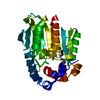
| ||||||||
|---|---|---|---|---|---|---|---|---|---|
| 1 |
| ||||||||
| Unit cell |
|
-Components
| #1: Protein | Mass: 35479.434 Da / Num. of mol.: 1 Source method: isolated from a genetically manipulated source Source: (gene. exp.) Francisella philomiragia subsp. philomiragia ATCC 25015 (bacteria)Gene: BZ13_192 / Production host: |
|---|---|
| #2: Water | ChemComp-HOH /  Mass: 18.015 Da / Num. of mol.: 216 / Source method: isolated from a natural source / Formula: H2O Mass: 18.015 Da / Num. of mol.: 216 / Source method: isolated from a natural source / Formula: H2O |
-Experimental details
-Experiment
| Experiment | Method: X-RAY DIFFRACTION / Number of used crystals: 1 |
|---|
- Sample preparation
| Crystal | Density Matthews: 2.14 Å3/Da / Density % sol: 42.51 % |
|---|---|
| Crystal grow | Temperature: 289.15 K / Method: counter-diffusion Details: 20% (v/v) PEG 6000, 100mM BICINE/sodium hydroxide (pH 9.0) |
-Data collection
| Diffraction | Mean temperature: 100 K / Serial crystal experiment: N |
|---|---|
| Diffraction source | Source: SYNCHROTRON / Site: SSRF  / Beamline: BL17U / Wavelength: 0.979 Å / Beamline: BL17U / Wavelength: 0.979 Å |
| Detector | Type: DECTRIS EIGER X 16M / Detector: PIXEL / Date: Apr 20, 2018 |
| Radiation | Protocol: MAD / Monochromatic (M) / Laue (L): M / Scattering type: x-ray |
| Radiation wavelength | Wavelength: 0.979 Å / Relative weight: 1 |
| Reflection | Resolution: 1.58→51.32 Å / Num. obs: 37190 / % possible obs: 91.5 % / Redundancy: 3 % / Net I/σ(I): 6.7 |
| Reflection shell | Resolution: 1.58→1.67 Å / Num. unique obs: 5609 |
- Processing
| Software |
| ||||||||||||||||||||||||||||||||||||||||||||||||||||||||||||||||||||||||||||||||||||||||||||||||||
|---|---|---|---|---|---|---|---|---|---|---|---|---|---|---|---|---|---|---|---|---|---|---|---|---|---|---|---|---|---|---|---|---|---|---|---|---|---|---|---|---|---|---|---|---|---|---|---|---|---|---|---|---|---|---|---|---|---|---|---|---|---|---|---|---|---|---|---|---|---|---|---|---|---|---|---|---|---|---|---|---|---|---|---|---|---|---|---|---|---|---|---|---|---|---|---|---|---|---|---|
| Refinement | Method to determine structure: MAD / Resolution: 1.584→40.208 Å / SU ML: 0.18 / Cross valid method: NONE / σ(F): 1.34 / Phase error: 25.41
| ||||||||||||||||||||||||||||||||||||||||||||||||||||||||||||||||||||||||||||||||||||||||||||||||||
| Solvent computation | Shrinkage radii: 0.9 Å / VDW probe radii: 1.11 Å | ||||||||||||||||||||||||||||||||||||||||||||||||||||||||||||||||||||||||||||||||||||||||||||||||||
| Refinement step | Cycle: LAST / Resolution: 1.584→40.208 Å
| ||||||||||||||||||||||||||||||||||||||||||||||||||||||||||||||||||||||||||||||||||||||||||||||||||
| Refine LS restraints |
| ||||||||||||||||||||||||||||||||||||||||||||||||||||||||||||||||||||||||||||||||||||||||||||||||||
| LS refinement shell |
|
