Movie
Movie Controller


+ Open data

- Basic information
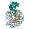
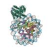
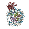
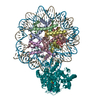
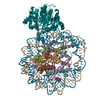
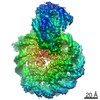
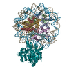
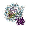
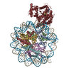
| Entry | Database: PDB / ID: 6k1p | |||||||||
|---|---|---|---|---|---|---|---|---|---|---|
| Title | The complex of ISWI-nucleosome in the ADP.BeF-bound state | |||||||||
 Components Components |
| |||||||||
 Keywords Keywords | DNA BINDING PROTEIN/DNA / chromatin remodelling / DNA BINDING PROTEIN-DNA complex | |||||||||
| Function / homology |  Function and homology information Function and homology informationIsw1b complex / Isw1a complex / Isw1 complex / regulation of transcriptional start site selection at RNA polymerase II promoter / nucleolar chromatin / negative regulation of RNA export from nucleus / negative regulation of IRE1-mediated unfolded protein response / regulation of chromatin organization / rDNA binding / nucleosome array spacer activity ...Isw1b complex / Isw1a complex / Isw1 complex / regulation of transcriptional start site selection at RNA polymerase II promoter / nucleolar chromatin / negative regulation of RNA export from nucleus / negative regulation of IRE1-mediated unfolded protein response / regulation of chromatin organization / rDNA binding / nucleosome array spacer activity / DNA-templated transcription elongation / sister chromatid cohesion / termination of RNA polymerase II transcription / termination of RNA polymerase I transcription / nucleosome binding / helicase activity / mRNA 3'-UTR binding / Hydrolases; Acting on acid anhydrides; Acting on acid anhydrides to facilitate cellular and subcellular movement / structural constituent of chromatin / nucleosome / nucleosome assembly / heterochromatin formation / response to heat / transcription cis-regulatory region binding / chromatin remodeling / protein heterodimerization activity / hydrolase activity / chromatin binding / regulation of DNA-templated transcription / chromatin / positive regulation of transcription by RNA polymerase II / DNA binding / ATP binding / nucleus Similarity search - Function | |||||||||
| Biological species |   | |||||||||
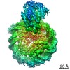
| Method | ELECTRON MICROSCOPY / single particle reconstruction / cryo EM / Resolution: 3.87 Å | |||||||||
 Authors Authors | Yan, L.J. / Wu, H. / Li, X.M. / Gao, N. / Chen, Z.C. | |||||||||
 Citation Citation | Journal: Nat Struct Mol Biol / Year: 2019 Title: Structures of the ISWI-nucleosome complex reveal a conserved mechanism of chromatin remodeling. Authors: Lijuan Yan / Hao Wu / Xuemei Li / Ning Gao / Zhucheng Chen /  Abstract: Chromatin remodelers are diverse enzymes, and different models have been proposed to explain how these proteins work. Here we report the 3.3 Å-resolution cryogenic electron microscopy (cryo-EM) ...Chromatin remodelers are diverse enzymes, and different models have been proposed to explain how these proteins work. Here we report the 3.3 Å-resolution cryogenic electron microscopy (cryo-EM) structures of Saccharomyces cerevisiae ISWI (ISW1) in complex with the nucleosome in adenosine diphosphate (ADP)-bound and ADP-BeF-bound states. The data show that after nucleosome binding, ISW1 is activated by substantial rearrangement of the catalytic domains, with the regulatory AutoN domain packing the first RecA-like core and the NegC domain being disordered. The high-resolution structure reveals local DNA distortion and translocation induced by ISW1 in the ADP-bound state, which is essentially identical to that induced by the Snf2 chromatin remodeler, suggesting a common mechanism of DNA translocation. The histone core remains largely unperturbed, and prevention of histone distortion by crosslinking did not inhibit the activity of yeast ISW1 or its human homolog. Together, our findings suggest a general mechanism of chromatin remodeling involving local DNA distortion without notable histone deformation. | |||||||||
| History |
|
- Structure visualization
| Movie |
Movie viewer |
|---|---|
| Structure viewer | Molecule: MolmilJmol/JSmol |
- Downloads & links
-Download
| PDBx/mmCIF format | 6k1p.cif.gz | 385.8 KB | Display | PDBx/mmCIF format |
|---|---|---|---|---|
| PDB format | pdb6k1p.ent.gz | 285.3 KB | Display | PDB format |
| PDBx/mmJSON format | 6k1p.json.gz | Tree view | PDBx/mmJSON format | |
| Others |  Other downloads Other downloads |
-Validation report
| Arichive directory | https://data.pdbj.org/pub/pdb/validation_reports/k1/6k1pftp://data.pdbj.org/pub/pdb/validation_reports/k1/6k1p | HTTPS FTP |
|---|
-Related structure data
| Related structure data | 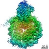 9719MC 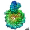 9718C 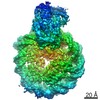 9720C  6iroC 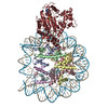 6jylC M: map data used to model this data C: citing same article ( |
|---|---|
| Similar structure data |
-Links
 PDBj
PDBj


- Assembly
| Deposited unit | 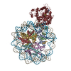
|
|---|---|
| 1 |
|
-Components
-Protein , 5 types, 9 molecules AEBFCGDHK
| #1: Protein | Mass: 15303.930 Da / Num. of mol.: 2 Source method: isolated from a genetically manipulated source Source: (gene. exp.) #2: Protein | Mass: 11263.231 Da / Num. of mol.: 2 Source method: isolated from a genetically manipulated source Source: (gene. exp.) #3: Protein | Mass: 13978.241 Da / Num. of mol.: 2 Source method: isolated from a genetically manipulated source Source: (gene. exp.) #4: Protein | Mass: 13524.752 Da / Num. of mol.: 2 Source method: isolated from a genetically manipulated source Source: (gene. exp.) #7: Protein | | Mass: 123170.516 Da / Num. of mol.: 1 Source method: isolated from a genetically manipulated source Source: (gene. exp.) Strain: ATCC 204508 / S288c / Gene: ISW1, YBR245C, YBR1633 / Production host: References: UniProt: P38144, Hydrolases; Acting on acid anhydrides; Acting on acid anhydrides to facilitate cellular and subcellular movement |
|---|
-DNA chain , 2 types, 2 molecules IJ
| #5: DNA chain | Mass: 51357.734 Da / Num. of mol.: 1 Source method: isolated from a genetically manipulated source Source: (gene. exp.) |
|---|---|
| #6: DNA chain | Mass: 51748.965 Da / Num. of mol.: 1 Source method: isolated from a genetically manipulated source Source: (gene. exp.) |
-Non-polymers , 3 types, 3 molecules 


| #8: Chemical | ChemComp-BEF /  Mass: 66.007 Da / Num. of mol.: 1 / Source method: obtained synthetically / Formula: BeF3 / Feature type: SUBJECT OF INVESTIGATION Mass: 66.007 Da / Num. of mol.: 1 / Source method: obtained synthetically / Formula: BeF3 / Feature type: SUBJECT OF INVESTIGATION |
|---|---|
| #9: Chemical | ChemComp-MG /  Mass: 24.305 Da / Num. of mol.: 1 / Source method: obtained synthetically / Formula: Mg / Feature type: SUBJECT OF INVESTIGATION Mass: 24.305 Da / Num. of mol.: 1 / Source method: obtained synthetically / Formula: Mg / Feature type: SUBJECT OF INVESTIGATION |
| #10: Chemical | ChemComp-ADP /  Mass: 427.201 Da / Num. of mol.: 1 / Source method: obtained synthetically / Formula: C10H15N5O10P2 / Feature type: SUBJECT OF INVESTIGATION / Comment: ADP, energy-carrying molecule*YM Mass: 427.201 Da / Num. of mol.: 1 / Source method: obtained synthetically / Formula: C10H15N5O10P2 / Feature type: SUBJECT OF INVESTIGATION / Comment: ADP, energy-carrying molecule*YM |
-Experimental details
-Experiment
| Experiment | Method: ELECTRON MICROSCOPY |
|---|---|
| EM experiment | Aggregation state: PARTICLE / 3D reconstruction method: single particle reconstruction |
- Sample preparation
| Component |
| ||||||||||||||||||||||||||||||
|---|---|---|---|---|---|---|---|---|---|---|---|---|---|---|---|---|---|---|---|---|---|---|---|---|---|---|---|---|---|---|---|
| Source (natural) |
| ||||||||||||||||||||||||||||||
| Source (recombinant) |
| ||||||||||||||||||||||||||||||
| Buffer solution | pH: 8.5 / Details: 10 mM Tris, 50 mM NaCl | ||||||||||||||||||||||||||||||
| Buffer component | Conc.: 1.35 mg/ml / Name: sodium chloride / Formula: NaCl | ||||||||||||||||||||||||||||||
| Specimen | Conc.: 0.1 mg/ml / Embedding applied: NO / Shadowing applied: NO / Staining applied: NO / Vitrification applied: YES / Details: monodisperse | ||||||||||||||||||||||||||||||
| Specimen support | Details: no special treatment / Grid material: COPPER / Grid mesh size: 400 divisions/in. / Grid type: Quantifoil R1.2/1.3 | ||||||||||||||||||||||||||||||
| Vitrification | Instrument: FEI VITROBOT MARK IV / Cryogen name: ETHANE / Humidity: 100 % / Chamber temperature: 281.15 K / Details: blot for 1.5s |
- Electron microscopy imaging
| Microscopy | Model: FEI TITAN / Details: performed manually |
|---|---|
| Electron gun | Electron source:  FIELD EMISSION GUN / Accelerating voltage: 300 kV / Illumination mode: SPOT SCAN FIELD EMISSION GUN / Accelerating voltage: 300 kV / Illumination mode: SPOT SCAN |
| Electron lens | Mode: DIFFRACTION / Nominal magnification: 5000 X / Calibrated magnification: 29000 X / Nominal defocus max: 5000 nm / Nominal defocus min: 1200 nm / Calibrated defocus min: 1200 nm / Calibrated defocus max: 5000 nm / Cs: 2.7 mm / C2 aperture diameter: 70 µm / Alignment procedure: BASIC |
| Specimen holder | Cryogen: HELIUM Specimen holder model: GATAN CT3500 SINGLE TILT LIQUID NITROGEN CRYO TRANSFER HOLDER Temperature (max): 100 K / Temperature (min): 100 K |
| Image recording | Electron dose: 1.5 e/Å2 / Film or detector model: GATAN K2 QUANTUM (4k x 4k) |
- Processing
| EM software |
| ||||||||||||||||||||||||||||||||||||||||
|---|---|---|---|---|---|---|---|---|---|---|---|---|---|---|---|---|---|---|---|---|---|---|---|---|---|---|---|---|---|---|---|---|---|---|---|---|---|---|---|---|---|
| CTF correction | Type: PHASE FLIPPING AND AMPLITUDE CORRECTION | ||||||||||||||||||||||||||||||||||||||||
| 3D reconstruction | Resolution: 3.87 Å / Resolution method: FSC 0.143 CUT-OFF / Num. of particles: 62121 / Algorithm: FOURIER SPACE / Details: relion / Num. of class averages: 5 / Symmetry type: POINT | ||||||||||||||||||||||||||||||||||||||||
| Atomic model building | B value: 200 / Protocol: AB INITIO MODEL / Space: REAL / Target criteria: correlation coefficient | ||||||||||||||||||||||||||||||||||||||||
| Atomic model building | PDB-ID: 5X0Y Pdb chain-ID: A / Accession code: 5X0Y / Details: chimera / Pdb chain residue range: 1-1000 / Source name: PDB / Type: experimental model |