 Movie
Movie Controller


+ Open data

- Basic information
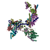
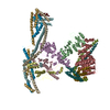
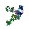
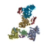
| Entry | Database: PDB / ID: 6k15 | ||||||
|---|---|---|---|---|---|---|---|
| Title | RSC substrate-recruitment module | ||||||
 Components Components |
| ||||||
 Keywords Keywords | DNA BINDING PROTEIN / chromatin remodeler / SWI/SNF family | ||||||
| Function / homology |  Function and homology information Function and homology informationregulation of sporulation resulting in formation of a cellular spore / histone H2A reader activity / : / : / : / regulation of nuclear cell cycle DNA replication / histone H3 reader activity / plasmid maintenance / CENP-A containing chromatin assembly / DNA translocase activity ...regulation of sporulation resulting in formation of a cellular spore / histone H2A reader activity / : / : / : / regulation of nuclear cell cycle DNA replication / histone H3 reader activity / plasmid maintenance / CENP-A containing chromatin assembly / DNA translocase activity / histone H3K14ac reader activity / nucleosome array spacer activity / RSC-type complex / nucleosome disassembly / ATP-dependent chromatin remodeler activity / SWI/SNF complex / sister chromatid cohesion / UV-damage excision repair / sporulation resulting in formation of a cellular spore / nuclear chromosome / rRNA transcription / histone H4 reader activity / chromosome, centromeric region / nucleosome binding / cytoskeleton organization / DNA helicase activity / helicase activity / transcription coregulator activity / positive regulation of transcription elongation by RNA polymerase II / chromosome segregation / transcription elongation by RNA polymerase II / meiotic cell cycle / chromatin DNA binding / base-excision repair / double-strand break repair via homologous recombination / double-strand break repair via nonhomologous end joining / G2/M transition of mitotic cell cycle / double-strand break repair / heterochromatin formation / histone binding / sequence-specific DNA binding / DNA helicase / DNA-binding transcription factor activity, RNA polymerase II-specific / chromatin remodeling / chromatin binding / regulation of transcription by RNA polymerase II / regulation of DNA-templated transcription / positive regulation of DNA-templated transcription / chromatin / structural molecule activity / positive regulation of transcription by RNA polymerase II / ATP hydrolysis activity / DNA binding / zinc ion binding / ATP binding / nucleus Similarity search - Function | ||||||
| Biological species |  | ||||||
| Method | ELECTRON MICROSCOPY / single particle reconstruction / cryo EM / Resolution: 3.4 Å | ||||||
 Authors Authors | Ye, Y.P. / Wu, H. / Chen, K.J. / Verma, N. / Cairns, B. / Gao, N. / Chen, Z.C. | ||||||
 Citation Citation | Journal: Science / Year: 2019 Title: Structure of the RSC complex bound to the nucleosome. Authors: Youpi Ye / Hao Wu / Kangjing Chen / Cedric R Clapier / Naveen Verma / Wenhao Zhang / Haiteng Deng / Bradley R Cairns / Ning Gao / Zhucheng Chen /   Abstract: The RSC complex remodels chromatin structure and regulates gene transcription. We used cryo-electron microscopy to determine the structure of yeast RSC bound to the nucleosome. RSC is delineated into ...The RSC complex remodels chromatin structure and regulates gene transcription. We used cryo-electron microscopy to determine the structure of yeast RSC bound to the nucleosome. RSC is delineated into the adenosine triphosphatase motor, the actin-related protein module, and the substrate recruitment module (SRM). RSC binds the nucleosome mainly through the motor, with the auxiliary subunit Sfh1 engaging the H2A-H2B acidic patch to enable nucleosome ejection. SRM is organized into three substrate-binding lobes poised to bind their respective nucleosomal epitopes. The relative orientations of the SRM and the motor on the nucleosome explain the directionality of DNA translocation and promoter nucleosome repositioning by RSC. Our findings shed light on RSC assembly and functionality, and they provide a framework to understand the mammalian homologs BAF/PBAF and the Sfh1 ortholog INI1/BAF47, which are frequently mutated in cancers. | ||||||
| History |
|
- Structure visualization
| Movie |
Movie viewer |
|---|---|
| Structure viewer | Molecule: MolmilJmol/JSmol |
- Downloads & links
-Download
| PDBx/mmCIF format | 6k15.cif.gz | 591.7 KB | Display | PDBx/mmCIF format |
|---|---|---|---|---|
| PDB format | pdb6k15.ent.gz | 414.8 KB | Display | PDB format |
| PDBx/mmJSON format | 6k15.json.gz | Tree view | PDBx/mmJSON format | |
| Others |  Other downloads Other downloads |
-Validation report
| Arichive directory | https://data.pdbj.org/pub/pdb/validation_reports/k1/6k15ftp://data.pdbj.org/pub/pdb/validation_reports/k1/6k15 | HTTPS FTP |
|---|
-Related structure data
| Related structure data |  9905MC  0777C  0778C  6kw3C  6kw4C M: map data used to model this data C: citing same article ( |
|---|---|
| Similar structure data |


-Links
 PDBj
PDBj


- Assembly
| Deposited unit | 
|
|---|---|
| 1 |
|
-Components
-Chromatin structure-remodeling complex subunit ... , 5 types, 5 molecules FMGXL
| #1: Protein | Mass: 49716.520 Da / Num. of mol.: 1 / Source method: isolated from a natural source / Source: (natural) |
|---|---|
| #3: Protein | Mass: 65289.309 Da / Num. of mol.: 1 / Source method: isolated from a natural source / Source: (natural) |
| #5: Protein | Mass: 48833.180 Da / Num. of mol.: 1 / Source method: isolated from a natural source / Source: (natural) |
| #11: Protein | Mass: 72372.375 Da / Num. of mol.: 1 / Source method: isolated from a natural source / Source: (natural) |
| #12: Protein | Mass: 102443.664 Da / Num. of mol.: 1 / Source method: isolated from a natural source / Source: (natural) |
-Chromatin structure-remodeling complex protein ... , 5 types, 6 molecules HDIACK
| #2: Protein | Mass: 63253.965 Da / Num. of mol.: 2 / Source method: isolated from a natural source / Source: (natural) #4: Protein | | Mass: 54222.691 Da / Num. of mol.: 1 / Source method: isolated from a natural source / Source: (natural) #6: Protein | | Mass: 57871.309 Da / Num. of mol.: 1 / Source method: isolated from a natural source / Source: (natural) #9: Protein | | Mass: 101448.211 Da / Num. of mol.: 1 / Source method: isolated from a natural source / Source: (natural) #10: Protein | | Mass: 101833.961 Da / Num. of mol.: 1 / Source method: isolated from a natural source / Source: (natural) |
|---|
-Protein , 2 types, 2 molecules JE
| #7: Protein | Mass: 156982.406 Da / Num. of mol.: 1 / Source method: isolated from a natural source / Source: (natural) |
|---|---|
| #8: Protein | Mass: 9192.524 Da / Num. of mol.: 1 / Source method: isolated from a natural source / Source: (natural) |
-Non-polymers , 1 types, 1 molecules 
| #13: Chemical | ChemComp-ZN /  Mass: 65.409 Da / Num. of mol.: 1 / Source method: obtained synthetically / Formula: Zn / Feature type: SUBJECT OF INVESTIGATION Mass: 65.409 Da / Num. of mol.: 1 / Source method: obtained synthetically / Formula: Zn / Feature type: SUBJECT OF INVESTIGATION |
|---|
-Details
| Has ligand of interest | Y |
|---|
-Experimental details
-Experiment
| Experiment | Method: ELECTRON MICROSCOPY |
|---|---|
| EM experiment | Aggregation state: CELL / 3D reconstruction method: single particle reconstruction |
- Sample preparation
| Component | Name: RSC / Type: COMPLEX / Entity ID: #1-#12 / Source: NATURAL |
|---|---|
| Source (natural) | Organism: |
| Buffer solution | pH: 7.5 |
| Specimen | Embedding applied: NO / Shadowing applied: NO / Staining applied: NO / Vitrification applied: YES |
| Vitrification | Cryogen name: ETHANE |
- Electron microscopy imaging
| Experimental equipment |  Model: Titan Krios / Image courtesy: FEI Company |
|---|---|
| Microscopy | Model: FEI TITAN KRIOS |
| Electron gun | Electron source:  FIELD EMISSION GUN / Accelerating voltage: 300 kV / Illumination mode: SPOT SCAN FIELD EMISSION GUN / Accelerating voltage: 300 kV / Illumination mode: SPOT SCAN |
| Electron lens | Mode: DARK FIELD |
| Image recording | Electron dose: 2 e/Å2 / Film or detector model: GATAN K2 QUANTUM (4k x 4k) |
- Processing
| CTF correction | Type: PHASE FLIPPING AND AMPLITUDE CORRECTION |
|---|---|
| 3D reconstruction | Resolution: 3.4 Å / Resolution method: FSC 0.143 CUT-OFF / Num. of particles: 280000 / Symmetry type: POINT |
