 Movie
Movie Controller

[English] 日本語
 Yorodumi
- PDB-6k0g: Crystal Structure of UDP-glucose 4-epimerase from Bifidobacterium... -
+ Open data

- Basic information
| Entry | Database: PDB / ID: 6k0g | ||||||
|---|---|---|---|---|---|---|---|
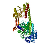
| Title | Crystal Structure of UDP-glucose 4-epimerase from Bifidobacterium longum in complex with NAD+ and UDP | ||||||
 Components Components | UDP-glucose 4-epimerase | ||||||
 Keywords Keywords | ISOMERASE / Rossmann fold | ||||||
| Function / homology |  Function and homology information Function and homology informationUDP-glucose 4-epimerase / UDP-glucose 4-epimerase activity / galactose metabolic process / carbohydrate metabolic process / cytosol Similarity search - Function | ||||||
| Biological species |  Bifidobacterium longum subsp. longum (bacteria) Bifidobacterium longum subsp. longum (bacteria) | ||||||
| Method |  X-RAY DIFFRACTION / SYNCHROTRON / MOLECULAR REPLACEMENT / Resolution: 1.8 Å X-RAY DIFFRACTION / SYNCHROTRON / MOLECULAR REPLACEMENT / Resolution: 1.8 Å | ||||||
 Authors Authors | Nam, Y.-W. / Nishimoto, M. / Arakawa, T. / Kitaoka, M. / Fushinobu, S. | ||||||
 Citation Citation | Journal: Sci Rep / Year: 2019 Title: Structural basis for broad substrate specificity of UDP-glucose 4-epimerase in the human milk oligosaccharide catabolic pathway of Bifidobacterium longum. Authors: Nam, Y.W. / Nishimoto, M. / Arakawa, T. / Kitaoka, M. / Fushinobu, S. | ||||||
| History |
|
- Structure visualization
| Structure viewer | Molecule: MolmilJmol/JSmol |
|---|
- Downloads & links
-Download
| PDBx/mmCIF format | 6k0g.cif.gz | 92.7 KB | Display | PDBx/mmCIF format |
|---|---|---|---|---|
| PDB format | pdb6k0g.ent.gz | 66.1 KB | Display | PDB format |
| PDBx/mmJSON format | 6k0g.json.gz | Tree view | PDBx/mmJSON format | |
| Others |  Other downloads Other downloads |
-Validation report
| Arichive directory | https://data.pdbj.org/pub/pdb/validation_reports/k0/6k0gftp://data.pdbj.org/pub/pdb/validation_reports/k0/6k0g | HTTPS FTP |
|---|
-Related structure data
| Related structure data |  6k0hC  6k0iC  1ek6S S: Starting model for refinement C: citing same article ( |
|---|---|
| Similar structure data |


-Links
 PDBj
PDBj


- Assembly
| Deposited unit | 
| ||||||||
|---|---|---|---|---|---|---|---|---|---|
| 1 | 
| ||||||||
| Unit cell |
|
-Components
| #1: Protein | Mass: 38341.117 Da / Num. of mol.: 1 Source method: isolated from a genetically manipulated source Source: (gene. exp.) Bifidobacterium longum subsp. longum (strain ATCC 15707 / DSM 20219 / JCM 1217 / NCTC 11818 / E194b) (bacteria)Strain: ATCC 15707 / DSM 20219 / JCM 1217 / NCTC 11818 / E194b Gene: lnpD, BLLJ_1620 / Variant: JCM 1217 / Plasmid: pET-30 / Production host: |
|---|---|
| #2: Chemical | ChemComp-UDP /   Type: RNA linking / Mass: 404.161 Da / Num. of mol.: 1 / Source method: obtained synthetically / Formula: C9H14N2O12P2 / Feature type: SUBJECT OF INVESTIGATION / Comment: UDP*YM Type: RNA linking / Mass: 404.161 Da / Num. of mol.: 1 / Source method: obtained synthetically / Formula: C9H14N2O12P2 / Feature type: SUBJECT OF INVESTIGATION / Comment: UDP*YM |
| #3: Chemical | ChemComp-NAD /   Mass: 663.425 Da / Num. of mol.: 1 / Source method: obtained synthetically / Formula: C21H27N7O14P2 / Feature type: SUBJECT OF INVESTIGATION / Comment: NAD*YM Mass: 663.425 Da / Num. of mol.: 1 / Source method: obtained synthetically / Formula: C21H27N7O14P2 / Feature type: SUBJECT OF INVESTIGATION / Comment: NAD*YM |
| #4: Chemical | ChemComp-MG /   Mass: 24.305 Da / Num. of mol.: 1 / Source method: obtained synthetically / Formula: Mg Mass: 24.305 Da / Num. of mol.: 1 / Source method: obtained synthetically / Formula: Mg |
| #5: Water | ChemComp-HOH /  Mass: 18.015 Da / Num. of mol.: 375 / Source method: isolated from a natural source / Formula: H2O Mass: 18.015 Da / Num. of mol.: 375 / Source method: isolated from a natural source / Formula: H2O |
-Experimental details
-Experiment
| Experiment | Method: X-RAY DIFFRACTION / Number of used crystals: 1 |
|---|
- Sample preparation
| Crystal | Density Matthews: 3.06 Å3/Da / Density % sol: 59.83 % |
|---|---|
| Crystal grow | Temperature: 277.15 K / Method: vapor diffusion, sitting drop / pH: 7.5 Details: 30% (v/v) PEG 400, 0.2 M MgCl, 0.1 M HEPES-NaOH (pH 7.5), 10 mM UDP |
-Data collection
| Diffraction | Mean temperature: 100 K / Serial crystal experiment: N |
|---|---|
| Diffraction source | Source: SYNCHROTRON / Site: Photon Factory  / Beamline: BL-17A / Wavelength: 0.979 Å / Beamline: BL-17A / Wavelength: 0.979 Å |
| Detector | Type: ADSC QUANTUM 270 / Detector: CCD / Date: May 5, 2013 |
| Radiation | Monochromator: Numerical link type Si(111) double crystal / Protocol: SINGLE WAVELENGTH / Monochromatic (M) / Laue (L): M / Scattering type: x-ray |
| Radiation wavelength | Wavelength: 0.979 Å / Relative weight: 1 |
| Reflection | Resolution: 1.8→50 Å / Num. obs: 44804 / % possible obs: 100 % / Redundancy: 18.1 % / Rmerge(I) obs: 0.122 / Net I/σ(I): 31.8 |
| Reflection shell | Resolution: 1.8→1.83 Å / Redundancy: 20.6 % / Rmerge(I) obs: 0.573 / Mean I/σ(I) obs: 6.7 / Num. unique obs: 2104 / % possible all: 100 |
- Processing
| Software |
| ||||||||||||||||||||||||||||||||||||||||||||||||||||||||||||
|---|---|---|---|---|---|---|---|---|---|---|---|---|---|---|---|---|---|---|---|---|---|---|---|---|---|---|---|---|---|---|---|---|---|---|---|---|---|---|---|---|---|---|---|---|---|---|---|---|---|---|---|---|---|---|---|---|---|---|---|---|---|
| Refinement | Method to determine structure: MOLECULAR REPLACEMENT Starting model: 1EK6 Resolution: 1.8→40.17 Å / Cor.coef. Fo:Fc: 0.967 / Cor.coef. Fo:Fc free: 0.958 / SU B: 1.697 / SU ML: 0.053 / Cross valid method: THROUGHOUT / σ(F): 0 / ESU R: 0.086 / ESU R Free: 0.088 Details: HYDROGENS HAVE BEEN ADDED IN THE RIDING POSITIONS U VALUES : REFINED INDIVIDUALLY
| ||||||||||||||||||||||||||||||||||||||||||||||||||||||||||||
| Solvent computation | Ion probe radii: 0.8 Å / Shrinkage radii: 0.8 Å / VDW probe radii: 1.2 Å | ||||||||||||||||||||||||||||||||||||||||||||||||||||||||||||
| Displacement parameters | Biso max: 72.58 Å2 / Biso mean: 21.326 Å2 / Biso min: 6.63 Å2
| ||||||||||||||||||||||||||||||||||||||||||||||||||||||||||||
| Refinement step | Cycle: final / Resolution: 1.8→40.17 Å
| ||||||||||||||||||||||||||||||||||||||||||||||||||||||||||||
| Refine LS restraints |
| ||||||||||||||||||||||||||||||||||||||||||||||||||||||||||||
| LS refinement shell | Resolution: 1.801→1.848 Å / Rfactor Rfree error: 0 / Total num. of bins used: 20
|
