 Movie
Movie Controller

[English] 日本語
 Yorodumi
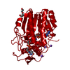
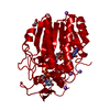
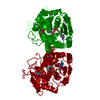
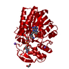
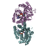
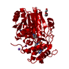
- PDB-1a9y: UDP-GALACTOSE 4-EPIMERASE MUTANT S124A/Y149F COMPLEXED WITH UDP-G... -
+ Open data

- Basic information
| Entry | Database: PDB / ID: 1a9y | |||||||||
|---|---|---|---|---|---|---|---|---|---|---|
| Title | UDP-GALACTOSE 4-EPIMERASE MUTANT S124A/Y149F COMPLEXED WITH UDP-GLUCOSE | |||||||||
 Components Components | UDP-GALACTOSE 4-EPIMERASE | |||||||||
 Keywords Keywords | EPIMERASE / GALACTOSE METABOLISM / DEHYDROGENASE | |||||||||
| Function / homology |  Function and homology information Function and homology informationmonosaccharide metabolic process / UDP-glucose 4-epimerase / colanic acid biosynthetic process / UDP-glucose 4-epimerase activity / racemase and epimerase activity, acting on carbohydrates and derivatives / galactose metabolic process / beta-D-galactose catabolic process via UDP-galactose, Leloir pathway / NAD+ binding / carbohydrate metabolic process / identical protein binding ...monosaccharide metabolic process / UDP-glucose 4-epimerase / colanic acid biosynthetic process / UDP-glucose 4-epimerase activity / racemase and epimerase activity, acting on carbohydrates and derivatives / galactose metabolic process / beta-D-galactose catabolic process via UDP-galactose, Leloir pathway / NAD+ binding / carbohydrate metabolic process / identical protein binding / cytosol / cytoplasm Similarity search - Function | |||||||||
| Biological species |  | |||||||||
| Method |  X-RAY DIFFRACTION / FOURIER DIFFERENCE / Resolution: 1.8 Å X-RAY DIFFRACTION / FOURIER DIFFERENCE / Resolution: 1.8 Å | |||||||||
 Authors Authors | Thoden, J.B. / Holden, H.M. | |||||||||
 Citation Citation | Journal: Biochemistry / Year: 1998 Title: Dramatic differences in the binding of UDP-galactose and UDP-glucose to UDP-galactose 4-epimerase from Escherichia coli. Authors: Thoden, J.B. / Holden, H.M. | |||||||||
| History |
|
- Structure visualization
| Structure viewer | Molecule: MolmilJmol/JSmol |
|---|
- Downloads & links
-Download
| PDBx/mmCIF format | 1a9y.cif.gz | 98.7 KB | Display | PDBx/mmCIF format |
|---|---|---|---|---|
| PDB format | pdb1a9y.ent.gz | 72.4 KB | Display | PDB format |
| PDBx/mmJSON format | 1a9y.json.gz | Tree view | PDBx/mmJSON format | |
| Others |  Other downloads Other downloads |
-Validation report
| Arichive directory | https://data.pdbj.org/pub/pdb/validation_reports/a9/1a9yftp://data.pdbj.org/pub/pdb/validation_reports/a9/1a9y | HTTPS FTP |
|---|
-Related structure data


-Links
 PDBj
PDBj
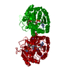
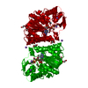
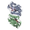
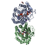
- Assembly
| Deposited unit | 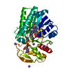
| |||||||||
|---|---|---|---|---|---|---|---|---|---|---|
| 1 | 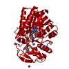
| |||||||||
| Unit cell |
| |||||||||
| Components on special symmetry positions |
|
-Components
| #1: Protein | Mass: 37276.043 Da / Num. of mol.: 1 / Mutation: S124A, Y149F Source method: isolated from a genetically manipulated source Source: (gene. exp.) | ||||||
|---|---|---|---|---|---|---|---|
| #2: Chemical | ChemComp-NA /   Mass: 22.990 Da / Num. of mol.: 4 / Source method: obtained synthetically / Formula: Na Mass: 22.990 Da / Num. of mol.: 4 / Source method: obtained synthetically / Formula: Na#3: Chemical | ChemComp-NAD / |   Mass: 663.425 Da / Num. of mol.: 1 / Source method: obtained synthetically / Formula: C21H27N7O14P2 / Comment: NAD*YM Mass: 663.425 Da / Num. of mol.: 1 / Source method: obtained synthetically / Formula: C21H27N7O14P2 / Comment: NAD*YM#4: Chemical | ChemComp-UPG / |   Mass: 566.302 Da / Num. of mol.: 1 / Source method: obtained synthetically / Formula: C15H24N2O17P2 Mass: 566.302 Da / Num. of mol.: 1 / Source method: obtained synthetically / Formula: C15H24N2O17P2#5: Water | ChemComp-HOH / |  Mass: 18.015 Da / Num. of mol.: 541 / Source method: isolated from a natural source / Formula: H2O Mass: 18.015 Da / Num. of mol.: 541 / Source method: isolated from a natural source / Formula: H2O |
-Experimental details
-Experiment
| Experiment | Method: X-RAY DIFFRACTION / Number of used crystals: 1 |
|---|
- Sample preparation
| Crystal | Density Matthews: 3 Å3/Da / Density % sol: 59 % | ||||||||||||||||||||||||||||||
|---|---|---|---|---|---|---|---|---|---|---|---|---|---|---|---|---|---|---|---|---|---|---|---|---|---|---|---|---|---|---|---|
| Crystal grow | pH: 9 Details: CRYSTALLIZED FROM 18% PEG8000, 500MM NACL, 20MM UDP-GLUCOSE THEN SOAKED IN 25% PEG8000, 750MM NACL, 20MM UDP-GLUCOSE, 20% ETHYLENE GLYCOL, pH 9.0 | ||||||||||||||||||||||||||||||
| Crystal | *PLUS | ||||||||||||||||||||||||||||||
| Crystal grow | *PLUS Temperature: 4 ℃ / Method: vapor diffusion, hanging drop | ||||||||||||||||||||||||||||||
| Components of the solutions | *PLUS
|
-Data collection
| Diffraction | Mean temperature: 120 K |
|---|---|
| Diffraction source | Source: ROTATING ANODE / Type: RIGAKU RUH2R / Wavelength: 1.5418 |
| Detector | Type: SIEMENS / Detector: AREA DETECTOR / Date: Mar 1, 1998 / Details: GOEBEL FOCUSING OPTICS |
| Radiation | Monochromator: NI FILTER / Monochromatic (M) / Laue (L): M / Scattering type: x-ray |
| Radiation wavelength | Wavelength: 1.5418 Å / Relative weight: 1 |
| Reflection | Resolution: 1.8→30 Å / Num. obs: 37966 / % possible obs: 91 % / Observed criterion σ(I): 0 / Redundancy: 2.1 % / Rmerge(I) obs: 0.041 / Rsym value: 0.041 / Net I/σ(I): 15 |
| Reflection shell | Resolution: 1.8→1.88 Å / Redundancy: 1.2 % / Rmerge(I) obs: 0.204 / Mean I/σ(I) obs: 1.9 / Rsym value: 0.204 / % possible all: 74 |
| Reflection | *PLUS Num. measured all: 84014 |
| Reflection shell | *PLUS % possible obs: 74 % / Num. unique obs: 3706 / Num. measured obs: 4531 |
- Processing
| Software |
| ||||||||||||||||||||||||||||||||||||||||||||||||||
|---|---|---|---|---|---|---|---|---|---|---|---|---|---|---|---|---|---|---|---|---|---|---|---|---|---|---|---|---|---|---|---|---|---|---|---|---|---|---|---|---|---|---|---|---|---|---|---|---|---|---|---|
| Refinement | Method to determine structure: FOURIER DIFFERENCE / Resolution: 1.8→30 Å / σ(F): 0 / Stereochemistry target values: TNT PROTGEO
| ||||||||||||||||||||||||||||||||||||||||||||||||||
| Solvent computation | Solvent model: TNT / Bsol: 689.4 Å2 / ksol: 1.011 e/Å3 | ||||||||||||||||||||||||||||||||||||||||||||||||||
| Refinement step | Cycle: LAST / Resolution: 1.8→30 Å
| ||||||||||||||||||||||||||||||||||||||||||||||||||
| Refine LS restraints |
| ||||||||||||||||||||||||||||||||||||||||||||||||||
| Software | *PLUS Name: TNT / Version: 5E / Classification: refinement | ||||||||||||||||||||||||||||||||||||||||||||||||||
| Refinement | *PLUS | ||||||||||||||||||||||||||||||||||||||||||||||||||
| Solvent computation | *PLUS | ||||||||||||||||||||||||||||||||||||||||||||||||||
| Displacement parameters | *PLUS | ||||||||||||||||||||||||||||||||||||||||||||||||||
| Refine LS restraints | *PLUS
|
