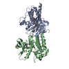
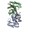
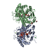
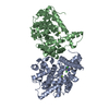
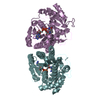
Entry Database : PDB / ID : 2c20Title CRYSTAL STRUCTURE OF UDP-GLUCOSE 4-EPIMERASE UDP-GLUCOSE 4-EPIMERASE Keywords / / / / Function / homology Function Domain/homology Component
/ / / / / / / / / / / / / / Biological species BACILLUS ANTHRACIS (anthrax bacterium)Method / / / Resolution : 2.7 Å Authors Lebedev, A.A. / Moroz, O.V. / Blagova, E.V. / Levdikov, V.M. / Fogg, M.J. / Brannigan, J.A. / Wilkinson, A.J. / Wilson, K.S. Journal : To be Published Title : Crystal Structure of Udp-Glucose 4-Epimerase from Bacillus Anthracis at 2.7A ResolutionAuthors : Lebedev, A.A. / Moroz, O.V. / Blagova, E.V. / Levdikov, V.M. / Fogg, M.J. / Brannigan, J.A. / Wilkinson, A.J. / Wilson, K.S. History Deposition Sep 22, 2005 Deposition site / Processing site Revision 1.0 Feb 20, 2007 Provider / Type Revision 1.1 May 8, 2011 Group Revision 1.2 Jul 13, 2011 Group Revision 1.3 Dec 13, 2023 Group Data collection / Database references ... Data collection / Database references / Derived calculations / Other / Refinement description Category chem_comp_atom / chem_comp_bond ... chem_comp_atom / chem_comp_bond / database_2 / pdbx_database_status / pdbx_initial_refinement_model / pdbx_struct_conn_angle / struct_conn / struct_site Item _database_2.pdbx_DOI / _database_2.pdbx_database_accession ... _database_2.pdbx_DOI / _database_2.pdbx_database_accession / _pdbx_database_status.status_code_sf / _pdbx_struct_conn_angle.ptnr1_auth_comp_id / _pdbx_struct_conn_angle.ptnr1_auth_seq_id / _pdbx_struct_conn_angle.ptnr1_label_atom_id / _pdbx_struct_conn_angle.ptnr1_label_comp_id / _pdbx_struct_conn_angle.ptnr1_label_seq_id / _pdbx_struct_conn_angle.ptnr3_auth_comp_id / _pdbx_struct_conn_angle.ptnr3_auth_seq_id / _pdbx_struct_conn_angle.ptnr3_label_atom_id / _pdbx_struct_conn_angle.ptnr3_label_comp_id / _pdbx_struct_conn_angle.ptnr3_label_seq_id / _pdbx_struct_conn_angle.value / _struct_conn.pdbx_dist_value / _struct_conn.pdbx_leaving_atom_flag / _struct_conn.ptnr1_auth_comp_id / _struct_conn.ptnr1_auth_seq_id / _struct_conn.ptnr1_label_asym_id / _struct_conn.ptnr1_label_atom_id / _struct_conn.ptnr1_label_comp_id / _struct_conn.ptnr1_label_seq_id / _struct_conn.ptnr2_auth_comp_id / _struct_conn.ptnr2_auth_seq_id / _struct_conn.ptnr2_label_asym_id / _struct_conn.ptnr2_label_atom_id / _struct_conn.ptnr2_label_comp_id / _struct_conn.ptnr2_label_seq_id / _struct_site.pdbx_auth_asym_id / _struct_site.pdbx_auth_comp_id / _struct_site.pdbx_auth_seq_id Revision 1.4 Oct 1, 2025 Group / Derived calculations / Structure summaryCategory pdbx_entry_details / pdbx_validate_close_contact ... pdbx_entry_details / pdbx_validate_close_contact / struct_conn / struct_conn_type Item
Show all Show less
 Movie
Movie Controller


 Open data
 Basic information
 Components
 Keywords
 Function and homology information
 X-RAY DIFFRACTION /
  Authors
 Citation
 Structure visualization
 Downloads & links
 Other downloads
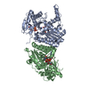


 PDBj
PDBj Assembly


 Mass: 663.425 Da / Num. of mol.: 6 / Source method: obtained synthetically / Formula: C21H27N7O14P2 / Comment: NAD*YM
 Mass: 65.409 Da / Num. of mol.: 6 / Source method: obtained synthetically / Formula: Zn
 Mass: 18.015 Da / Num. of mol.: 234 / Source method: isolated from a natural source / Formula: H2O
 Sample preparation
 / Beamline: BM14 / Wavelength: 1.00806
  Processing