 Movie
Movie Controller

[English] 日本語
 Yorodumi
- PDB-1lrl: Crystal Structure of UDP-Galactose 4-Epimerase Mutant Y299C Compl... -
+ Open data

- Basic information
| Entry | Database: PDB / ID: 1lrl | ||||||
|---|---|---|---|---|---|---|---|
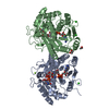
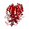
| Title | Crystal Structure of UDP-Galactose 4-Epimerase Mutant Y299C Complexed with UDP-Glucose | ||||||
 Components Components | UDP-glucose 4-epimerase | ||||||
 Keywords Keywords | ISOMERASE / epimerase / short chain dehydrogenase / galactosemia | ||||||
| Function / homology |  Function and homology information Function and homology informationmonosaccharide metabolic process / colanic acid biosynthetic process / UDP-glucose 4-epimerase / UDP-glucose 4-epimerase activity / racemase and epimerase activity, acting on carbohydrates and derivatives / beta-D-galactose catabolic process via UDP-galactose, Leloir pathway / galactose metabolic process / NAD+ binding / carbohydrate metabolic process / identical protein binding ...monosaccharide metabolic process / colanic acid biosynthetic process / UDP-glucose 4-epimerase / UDP-glucose 4-epimerase activity / racemase and epimerase activity, acting on carbohydrates and derivatives / beta-D-galactose catabolic process via UDP-galactose, Leloir pathway / galactose metabolic process / NAD+ binding / carbohydrate metabolic process / identical protein binding / cytoplasm / cytosol Similarity search - Function | ||||||
| Biological species |  | ||||||
| Method |  X-RAY DIFFRACTION / MOLECULAR REPLACEMENT / Resolution: 1.8 Å X-RAY DIFFRACTION / MOLECULAR REPLACEMENT / Resolution: 1.8 Å | ||||||
 Authors Authors | Thoden, J.B. / Henderson, J.M. / Fridovich-Keil, J.L. / Holden, H.M. | ||||||
 Citation Citation | Journal: J.Biol.Chem. / Year: 2002 Title: Structural analysis of the Y299C mutant of Escherichia coli UDP-galactose 4-epimerase. Teaching an old dog new tricks. Authors: Thoden, J.B. / Henderson, J.M. / Fridovich-Keil, J.L. / Holden, H.M. | ||||||
| History |
|
- Structure visualization
| Structure viewer | Molecule: MolmilJmol/JSmol |
|---|
- Downloads & links
-Download
| PDBx/mmCIF format | 1lrl.cif.gz | 93.6 KB | Display | PDBx/mmCIF format |
|---|---|---|---|---|
| PDB format | pdb1lrl.ent.gz | 69.3 KB | Display | PDB format |
| PDBx/mmJSON format | 1lrl.json.gz | Tree view | PDBx/mmJSON format | |
| Others |  Other downloads Other downloads |
-Validation report
| Arichive directory | https://data.pdbj.org/pub/pdb/validation_reports/lr/1lrlftp://data.pdbj.org/pub/pdb/validation_reports/lr/1lrl | HTTPS FTP |
|---|
-Related structure data
| Related structure data |  1lrjC  1lrkC  1ek6S C: citing same article ( S: Starting model for refinement |
|---|---|
| Similar structure data |


-Links
 PDBj
PDBj
- Assembly
| Deposited unit | 
| ||||||||||||||||||
|---|---|---|---|---|---|---|---|---|---|---|---|---|---|---|---|---|---|---|---|
| 1 | 
| ||||||||||||||||||
| Unit cell |
| ||||||||||||||||||
| Components on special symmetry positions |
| ||||||||||||||||||
| Details | The biological assembly is a homodimer. There is one independent monomer in the unit cell sitting on a crystallographic 2-fold axis. The matrix to generate the second subunit is: 1 0 0 0 -1 0 0 0 -1 0.0 0.0 36.5 |
-Components
-Protein , 1 types, 1 molecules A
| #1: Protein | Mass: 37233.984 Da / Num. of mol.: 1 / Mutation: Y299C Source method: isolated from a genetically manipulated source Source: (gene. exp.) |
|---|
-Non-polymers , 5 types, 397 molecules 


| #2: Chemical |  Mass: 22.990 Da / Num. of mol.: 2 / Source method: obtained synthetically / Formula: Na Mass: 22.990 Da / Num. of mol.: 2 / Source method: obtained synthetically / Formula: Na#3: Chemical | ChemComp-NAD / |  Mass: 663.425 Da / Num. of mol.: 1 / Source method: obtained synthetically / Formula: C21H27N7O14P2 / Comment: NAD*YM Mass: 663.425 Da / Num. of mol.: 1 / Source method: obtained synthetically / Formula: C21H27N7O14P2 / Comment: NAD*YM#4: Chemical | ChemComp-UPG / |  Mass: 566.302 Da / Num. of mol.: 1 / Source method: obtained synthetically / Formula: C15H24N2O17P2 Mass: 566.302 Da / Num. of mol.: 1 / Source method: obtained synthetically / Formula: C15H24N2O17P2#5: Chemical | ChemComp-PGE / |  Mass: 150.173 Da / Num. of mol.: 1 / Source method: obtained synthetically / Formula: C6H14O4 Mass: 150.173 Da / Num. of mol.: 1 / Source method: obtained synthetically / Formula: C6H14O4#6: Water | ChemComp-HOH / | Mass: 18.015 Da / Num. of mol.: 392 / Source method: isolated from a natural source / Formula: H2O |
|---|
-Experimental details
-Experiment
| Experiment | Method: X-RAY DIFFRACTION / Number of used crystals: 1 |
|---|
- Sample preparation
| Crystal | Density Matthews: 2.94 Å3/Da / Density % sol: 58.12 % | ||||||||||||||||||||||||||||||||||||||||||
|---|---|---|---|---|---|---|---|---|---|---|---|---|---|---|---|---|---|---|---|---|---|---|---|---|---|---|---|---|---|---|---|---|---|---|---|---|---|---|---|---|---|---|---|
| Crystal grow | Temperature: 277 K / Method: vapor diffusion, hanging drop / pH: 8 Details: PEG 8000, HEPPS, NaCl, UDP-glucose, pH 8.0, VAPOR DIFFUSION, HANGING DROP at 277K | ||||||||||||||||||||||||||||||||||||||||||
| Crystal grow | *PLUS Temperature: 4 ℃ | ||||||||||||||||||||||||||||||||||||||||||
| Components of the solutions | *PLUS
|
-Data collection
| Diffraction | Mean temperature: 115 K |
|---|---|
| Diffraction source | Source: ROTATING ANODE / Type: RIGAKU RU200 / Wavelength: 1.5418 Å |
| Detector | Type: SIEMENS HI-STAR / Detector: AREA DETECTOR / Date: Jan 21, 2002 / Details: Supper Mirrors |
| Radiation | Monochromator: Ni FILTER / Protocol: SINGLE WAVELENGTH / Monochromatic (M) / Laue (L): M / Scattering type: x-ray |
| Radiation wavelength | Wavelength: 1.5418 Å / Relative weight: 1 |
| Reflection | Resolution: 1.8→30 Å / Num. all: 39438 / Num. obs: 39438 / % possible obs: 93.1 % / Observed criterion σ(F): 0 / Observed criterion σ(I): 0 / Redundancy: 3.6 % / Rsym value: 0.054 / Net I/σ(I): 14.7 |
| Reflection shell | Resolution: 1.8→1.88 Å / Redundancy: 1.9 % / Mean I/σ(I) obs: 2.1 / Num. unique all: 4523 / Rsym value: 0.337 / % possible all: 82.5 |
| Reflection | *PLUS Rmerge(I) obs: 0.054 |
| Reflection shell | *PLUS % possible obs: 82.5 % / Num. unique obs: 4523 / Rmerge(I) obs: 0.337 |
- Processing
| Software |
| |||||||||||||||||||||||||
|---|---|---|---|---|---|---|---|---|---|---|---|---|---|---|---|---|---|---|---|---|---|---|---|---|---|---|
| Refinement | Method to determine structure: MOLECULAR REPLACEMENT Starting model: PDB entry 1EK6 Resolution: 1.8→30 Å / Cross valid method: THROUGHOUT / σ(F): 0 / Stereochemistry target values: Engh & Huber
| |||||||||||||||||||||||||
| Refinement step | Cycle: LAST / Resolution: 1.8→30 Å
| |||||||||||||||||||||||||
| Refine LS restraints |
| |||||||||||||||||||||||||
| Refinement | *PLUS Num. reflection obs: 35935 / Rfactor all: 0.195 / Rfactor Rfree: 0.261 / Rfactor Rwork: 0.194 | |||||||||||||||||||||||||
| Solvent computation | *PLUS | |||||||||||||||||||||||||
| Displacement parameters | *PLUS | |||||||||||||||||||||||||
| Refine LS restraints | *PLUS
|
