 Movie
Movie Controller

[English] 日本語
 Yorodumi
- PDB-6jzj: Structure of FimA type-2 (FimA2) prepilin of the type V major fimbrium -
+ Open data

- Basic information
| Entry | Database: PDB / ID: 6jzj | ||||||
|---|---|---|---|---|---|---|---|
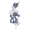
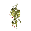
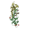
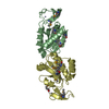
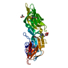
| Title | Structure of FimA type-2 (FimA2) prepilin of the type V major fimbrium | ||||||
 Components Components | Major fimbrium subunit FimA type-2 | ||||||
 Keywords Keywords | CELL ADHESION / Type V pili / Major fimbrial subunit protein | ||||||
| Function / homology |  Function and homology information Function and homology informationpilus / cell outer membrane / cell adhesion / structural molecule activity Similarity search - Function | ||||||
| Biological species |  Porphyromonas gingivalis TDC60 (bacteria) Porphyromonas gingivalis TDC60 (bacteria) | ||||||
| Method |  X-RAY DIFFRACTION / SYNCHROTRON / MOLECULAR REPLACEMENT / Resolution: 1.6 Å X-RAY DIFFRACTION / SYNCHROTRON / MOLECULAR REPLACEMENT / Resolution: 1.6 Å | ||||||
 Authors Authors | Okada, K. / Shoji, M. / Nakayam, K. / Imada, K. | ||||||
| Funding support |  Japan, 1items Japan, 1items
| ||||||
 Citation Citation | Journal: Nat Microbiol / Year: 2020 Title: Structure of polymerized type V pilin reveals assembly mechanism involving protease-mediated strand exchange. Authors: Satoshi Shibata / Mikio Shoji / Kodai Okada / Hideyuki Matsunami / Melissa M Matthews / Katsumi Imada / Koji Nakayama / Matthias Wolf / Abstract: Bacterial adhesion is a general strategy for host-microbe and microbe-microbe interactions. Adhesive pili are essential for colonization, biofilm formation, virulence and pathogenesis of many ...Bacterial adhesion is a general strategy for host-microbe and microbe-microbe interactions. Adhesive pili are essential for colonization, biofilm formation, virulence and pathogenesis of many environmental and pathogenic bacteria. Members of the class Bacteroidia have unique type V pili, assembled by protease-mediated polymerization. Porphyromonas gingivalis is the main contributor to periodontal disease and its type V pili are a key factor for its virulence. However, the structure of the polymerized pilus and its assembly mechanism are unknown. Here we show structures of polymerized and monomeric states of FimA stalk pilin from P. gingivalis, determined by cryo-electron microscopy and crystallography. The atomic model of assembled FimA shows that the C-terminal strand of a donor subunit is inserted into a groove in the β-sheet of an acceptor subunit after N-terminal cleavage by the protease RgpB. The C terminus of the donor strand is essential for polymerization. We propose that type V pili assemble via a sequential polar assembly mechanism at the cell surface, involving protease-mediated strand exchange, employed by various Gram-negative species belonging to the class Bacteroidia. Our results reveal functional surfaces related to pathogenic properties of polymerized FimA. These insights may facilitate development of antibacterial drugs. | ||||||
| History |
|
- Structure visualization
| Structure viewer | Molecule: MolmilJmol/JSmol |
|---|
- Downloads & links
-Download
| PDBx/mmCIF format | 6jzj.cif.gz | 90.8 KB | Display | PDBx/mmCIF format |
|---|---|---|---|---|
| PDB format | pdb6jzj.ent.gz | 65.1 KB | Display | PDB format |
| PDBx/mmJSON format | 6jzj.json.gz | Tree view | PDBx/mmJSON format | |
| Others |  Other downloads Other downloads |
-Validation report
| Arichive directory | https://data.pdbj.org/pub/pdb/validation_reports/jz/6jzjftp://data.pdbj.org/pub/pdb/validation_reports/jz/6jzj | HTTPS FTP |
|---|
-Related structure data
| Related structure data |  0724C 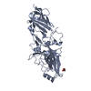 6jzkC  6kmfC  4q98S S: Starting model for refinement C: citing same article ( |
|---|---|
| Similar structure data |


-Links
 PDBj
PDBj


- Assembly
| Deposited unit | 
| ||||||||
|---|---|---|---|---|---|---|---|---|---|
| 1 |
| ||||||||
| Unit cell |
|
-Components
| #1: Protein | Mass: 40500.863 Da / Num. of mol.: 1 Source method: isolated from a genetically manipulated source Source: (gene. exp.) Porphyromonas gingivalis TDC60 (bacteria)Gene: fimA (type II) / Plasmid: pET15b / Production host: | ||
|---|---|---|---|
| #2: Chemical |   Mass: 96.063 Da / Num. of mol.: 2 / Source method: obtained synthetically / Formula: SO4 Mass: 96.063 Da / Num. of mol.: 2 / Source method: obtained synthetically / Formula: SO4#3: Water | ChemComp-HOH / |  Mass: 18.015 Da / Num. of mol.: 351 / Source method: isolated from a natural source / Formula: H2O Mass: 18.015 Da / Num. of mol.: 351 / Source method: isolated from a natural source / Formula: H2O |
-Experimental details
-Experiment
| Experiment | Method: X-RAY DIFFRACTION / Number of used crystals: 1 |
|---|
- Sample preparation
| Crystal | Density Matthews: 3.14 Å3/Da / Density % sol: 60.87 % |
|---|---|
| Crystal grow | Temperature: 293 K / Method: vapor diffusion, sitting drop / pH: 7.5 Details: 0.1M HEPES-NaOH pH 7.5, 2.2M ammonium sulfate, 1.5% PEG 400 |
-Data collection
| Diffraction | Mean temperature: 100 K / Serial crystal experiment: N |
|---|---|
| Diffraction source | Source: SYNCHROTRON / Site: SPring-8 / Beamline: BL41XU / Wavelength: 1 Å |
| Detector | Type: DECTRIS PILATUS3 6M / Detector: PIXEL / Date: Jun 18, 2017 |
| Radiation | Monochromator: Double-crystal / Protocol: SINGLE WAVELENGTH / Monochromatic (M) / Laue (L): M / Scattering type: x-ray |
| Radiation wavelength | Wavelength: 1 Å / Relative weight: 1 |
| Reflection | Resolution: 1.6→65.8 Å / Num. obs: 68085 / % possible obs: 99.9 % / Redundancy: 4.7 % / Biso Wilson estimate: 15.5 Å2 / CC1/2: 0.999 / Rmerge(I) obs: 0.051 / Rpim(I) all: 0.039 / Rrim(I) all: 0.064 / Net I/σ(I): 13.7 |
| Reflection shell | Resolution: 1.6→1.63 Å / Redundancy: 4.7 % / Rmerge(I) obs: 0.514 / Num. unique obs: 3344 / CC1/2: 0.837 / Rpim(I) all: 0.403 / Rrim(I) all: 0.656 / % possible all: 100 |
- Processing
| Software |
| ||||||||||||||||||||||||||||||||||||||||||||||||||||||||||||||||||||||||||||||||||||||||||||||||||||||||||||||||||||||||||||||||||||||||||||||||||||||||||||||||||||||||||||||||||||||
|---|---|---|---|---|---|---|---|---|---|---|---|---|---|---|---|---|---|---|---|---|---|---|---|---|---|---|---|---|---|---|---|---|---|---|---|---|---|---|---|---|---|---|---|---|---|---|---|---|---|---|---|---|---|---|---|---|---|---|---|---|---|---|---|---|---|---|---|---|---|---|---|---|---|---|---|---|---|---|---|---|---|---|---|---|---|---|---|---|---|---|---|---|---|---|---|---|---|---|---|---|---|---|---|---|---|---|---|---|---|---|---|---|---|---|---|---|---|---|---|---|---|---|---|---|---|---|---|---|---|---|---|---|---|---|---|---|---|---|---|---|---|---|---|---|---|---|---|---|---|---|---|---|---|---|---|---|---|---|---|---|---|---|---|---|---|---|---|---|---|---|---|---|---|---|---|---|---|---|---|---|---|---|---|
| Refinement | Method to determine structure: MOLECULAR REPLACEMENT Starting model: 4Q98 Resolution: 1.6→65.75 Å / SU ML: 0.17 / Cross valid method: THROUGHOUT / σ(F): 1.34 / Phase error: 19.5
| ||||||||||||||||||||||||||||||||||||||||||||||||||||||||||||||||||||||||||||||||||||||||||||||||||||||||||||||||||||||||||||||||||||||||||||||||||||||||||||||||||||||||||||||||||||||
| Solvent computation | Shrinkage radii: 0.9 Å / VDW probe radii: 1.11 Å | ||||||||||||||||||||||||||||||||||||||||||||||||||||||||||||||||||||||||||||||||||||||||||||||||||||||||||||||||||||||||||||||||||||||||||||||||||||||||||||||||||||||||||||||||||||||
| Refinement step | Cycle: LAST / Resolution: 1.6→65.75 Å
| ||||||||||||||||||||||||||||||||||||||||||||||||||||||||||||||||||||||||||||||||||||||||||||||||||||||||||||||||||||||||||||||||||||||||||||||||||||||||||||||||||||||||||||||||||||||
| Refine LS restraints |
| ||||||||||||||||||||||||||||||||||||||||||||||||||||||||||||||||||||||||||||||||||||||||||||||||||||||||||||||||||||||||||||||||||||||||||||||||||||||||||||||||||||||||||||||||||||||
| LS refinement shell |
|
