 Movie
Movie Controller


+ Open data

- Basic information
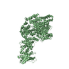
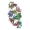
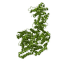
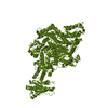
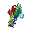
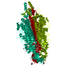
| Entry | Database: PDB / ID: 6jxr | |||||||||||||||||||||||||||||||||||||||||||||||||||||||||||||||||||||
|---|---|---|---|---|---|---|---|---|---|---|---|---|---|---|---|---|---|---|---|---|---|---|---|---|---|---|---|---|---|---|---|---|---|---|---|---|---|---|---|---|---|---|---|---|---|---|---|---|---|---|---|---|---|---|---|---|---|---|---|---|---|---|---|---|---|---|---|---|---|---|
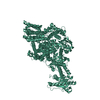
| Title | Structure of human T cell receptor-CD3 complex | |||||||||||||||||||||||||||||||||||||||||||||||||||||||||||||||||||||
 Components Components |
| |||||||||||||||||||||||||||||||||||||||||||||||||||||||||||||||||||||
 Keywords Keywords | IMMUNE SYSTEM | |||||||||||||||||||||||||||||||||||||||||||||||||||||||||||||||||||||
| Function / homology |  Function and homology information Function and homology informationregulation of lymphocyte apoptotic process / gamma-delta T cell receptor complex / Fc-gamma receptor III complex / T cell anergy / T cell activation involved in immune response / positive regulation of T cell anergy / CD4-positive, alpha-beta T cell proliferation / Fc-gamma receptor signaling pathway / gamma-delta T cell activation / positive regulation of cell-cell adhesion mediated by integrin ...regulation of lymphocyte apoptotic process / gamma-delta T cell receptor complex / Fc-gamma receptor III complex / T cell anergy / T cell activation involved in immune response / positive regulation of T cell anergy / CD4-positive, alpha-beta T cell proliferation / Fc-gamma receptor signaling pathway / gamma-delta T cell activation / positive regulation of cell-cell adhesion mediated by integrin / negative thymic T cell selection / positive regulation of CD4-positive, alpha-beta T cell proliferation / positive thymic T cell selection / signal complex assembly / positive regulation of protein localization to cell surface / Nef and signal transduction / alpha-beta T cell receptor complex / T cell receptor complex / positive regulation of cell-matrix adhesion / smoothened signaling pathway / Translocation of ZAP-70 to Immunological synapse / Phosphorylation of CD3 and TCR zeta chains / positive regulation of interleukin-4 production / dendrite development / protein complex oligomerization / establishment or maintenance of cell polarity / alpha-beta T cell activation / FCGR activation / Generation of second messenger molecules / immunological synapse / Co-inhibition by PD-1 / Role of phospholipids in phagocytosis / positive regulation of interleukin-2 production / T cell receptor binding / T cell costimulation / endomembrane system / positive regulation of T cell proliferation / positive regulation of calcium-mediated signaling / FCGR3A-mediated IL10 synthesis / cell surface receptor protein tyrosine kinase signaling pathway / cerebellum development / protein tyrosine kinase binding / T cell activation / FCGR3A-mediated phagocytosis / response to bacterium / negative regulation of smoothened signaling pathway / clathrin-coated endocytic vesicle membrane / apoptotic signaling pathway / calcium-mediated signaling / SH3 domain binding / Regulation of actin dynamics for phagocytic cup formation / peptide antigen binding / positive regulation of type II interferon production / Immunoregulatory interactions between a Lymphoid and a non-Lymphoid cell / cell-cell junction / transmembrane signaling receptor activity / Downstream TCR signaling / Cargo recognition for clathrin-mediated endocytosis / protein transport / T cell receptor signaling pathway / Clathrin-mediated endocytosis / signaling receptor complex adaptor activity / cell body / protein-containing complex assembly / regulation of apoptotic process / dendritic spine / adaptive immune response / protein-macromolecule adaptor activity / cell surface receptor signaling pathway / G protein-coupled receptor signaling pathway / protein heterodimerization activity / external side of plasma membrane / negative regulation of gene expression / positive regulation of gene expression / protein kinase binding / cell surface / Golgi apparatus / endoplasmic reticulum / protein homodimerization activity / identical protein binding / plasma membrane / cytoplasm / cytosol Similarity search - Function | |||||||||||||||||||||||||||||||||||||||||||||||||||||||||||||||||||||
| Biological species |  Homo sapiens (human) Homo sapiens (human) | |||||||||||||||||||||||||||||||||||||||||||||||||||||||||||||||||||||
| Method | ELECTRON MICROSCOPY / single particle reconstruction / negative staining / cryo EM / Resolution: 3.7 Å | |||||||||||||||||||||||||||||||||||||||||||||||||||||||||||||||||||||
 Authors Authors | Dong, D. / Zheng, L. / Lin, J. / Zhu, Y. / Li, N. / Zhang, B. / Xie, S. / Zheng, J. / Wang, Y. / Gao, N. / Huang, Z. | |||||||||||||||||||||||||||||||||||||||||||||||||||||||||||||||||||||
| Funding support |  China, 1items China, 1items
| |||||||||||||||||||||||||||||||||||||||||||||||||||||||||||||||||||||
 Citation Citation | Journal: Nature / Year: 2019 Title: Structural basis of assembly of the human T cell receptor-CD3 complex. Authors: De Dong / Lvqin Zheng / Jianquan Lin / Bailing Zhang / Yuwei Zhu / Ningning Li / Shuangyu Xie / Yuhang Wang / Ning Gao / Zhiwei Huang / Abstract: The αβ T cell receptor (TCR), in association with the CD3γε-CD3δε-CD3ζζ signalling hexamer, is the primary determinant of T cell development and activation, and of immune responses to foreign ...The αβ T cell receptor (TCR), in association with the CD3γε-CD3δε-CD3ζζ signalling hexamer, is the primary determinant of T cell development and activation, and of immune responses to foreign antigens. The mechanism of assembly of the TCR-CD3 complex remains unknown. Here we report a cryo-electron microscopy structure of human TCRαβ in complex with the CD3 hexamer at 3.7 Å resolution. The structure contains the complete extracellular domains and all the transmembrane helices of TCR-CD3. The octameric TCR-CD3 complex is assembled with 1:1:1:1 stoichiometry of TCRαβ:CD3γε:CD3δε:CD3ζζ. Assembly of the extracellular domains of TCR-CD3 is mediated by the constant domains and connecting peptides of TCRαβ that pack against CD3γε-CD3δε, forming a trimer-like structure proximal to the plasma membrane. The transmembrane segment of the CD3 complex adopts a barrel-like structure formed by interaction of the two transmembrane helices of CD3ζζ with those of CD3γε and CD3δε. Insertion of the transmembrane helices of TCRαβ into the barrel-like structure via both hydrophobic and ionic interactions results in transmembrane assembly of the TCR-CD3 complex. Together, our data reveal the structural basis for TCR-CD3 complex assembly, providing clues to TCR triggering and a foundation for rational design of immunotherapies that target the complex. | |||||||||||||||||||||||||||||||||||||||||||||||||||||||||||||||||||||
| History |
|
- Structure visualization
| Movie |
Movie viewer |
|---|---|
| Structure viewer | Molecule: MolmilJmol/JSmol |
- Downloads & links
-Download
| PDBx/mmCIF format | 6jxr.cif.gz | 209.1 KB | Display | PDBx/mmCIF format |
|---|---|---|---|---|
| PDB format | pdb6jxr.ent.gz | 159.4 KB | Display | PDB format |
| PDBx/mmJSON format | 6jxr.json.gz | Tree view | PDBx/mmJSON format | |
| Others |  Other downloads Other downloads |
-Validation report
| Arichive directory | https://data.pdbj.org/pub/pdb/validation_reports/jx/6jxrftp://data.pdbj.org/pub/pdb/validation_reports/jx/6jxr | HTTPS FTP |
|---|
-Related structure data
| Related structure data | 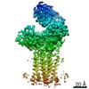 9895MC M: map data used to model this data C: citing same article ( |
|---|---|
| Similar structure data |
-Links
 PDBj
PDBj


- Assembly
| Deposited unit | 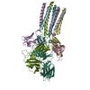
|
|---|---|
| 1 |
|
-Components
-T-cell surface glycoprotein CD3 ... , 4 types, 6 molecules abdfeg
| #1: Protein | Mass: 18723.439 Da / Num. of mol.: 2 Source method: isolated from a genetically manipulated source Source: (gene. exp.) Homo sapiens (human) / Gene: CD247, CD3Z, T3Z, TCRZ / Production host: Homo sapiens (human) / References: UniProt: P20963#2: Protein | | Mass: 18949.537 Da / Num. of mol.: 1 Source method: isolated from a genetically manipulated source Source: (gene. exp.) Homo sapiens (human) / Gene: CD3D, T3D / Production host: Homo sapiens (human) / References: UniProt: P04234#3: Protein | Mass: 23174.227 Da / Num. of mol.: 2 Source method: isolated from a genetically manipulated source Source: (gene. exp.) Homo sapiens (human) / Gene: CD3E, T3E / Production host: Homo sapiens (human) / References: UniProt: P07766#4: Protein | | Mass: 20493.457 Da / Num. of mol.: 1 Source method: isolated from a genetically manipulated source Source: (gene. exp.) Homo sapiens (human) / Gene: CD3G, T3G / Production host: Homo sapiens (human) / References: UniProt: P09693 |
|---|
-T cell receptor ... , 2 types, 2 molecules mn
| #5: Protein | Mass: 28308.662 Da / Num. of mol.: 1 Source method: isolated from a genetically manipulated source Details: fusion protein of residues 22-114 from A0A0B4J271, residues 116-132 from A0N4Z6 and residues 134-273 from P01848. Source: (gene. exp.) Homo sapiens (human) / Gene: TRAV12-3, Tcr-alpha, TRAC, TCRA / Production host: Homo sapiens (human)References: UniProt: A0A0B4J271, UniProt: A0N4Z6, UniProt: P01848 |
|---|---|
| #6: Protein | Mass: 32498.463 Da / Num. of mol.: 1 Source method: isolated from a genetically manipulated source Details: fusion protein of residues 22-112 from A0A0K0K1A5, residues 121-134 from P0DSE2 and residues 135-312 from A0A0G2JMB4. Source: (gene. exp.) Homo sapiens (human) / Gene: TRBV6-5, TRB, TRBC2 / Production host: Homo sapiens (human)References: UniProt: A0A0K0K1A5, UniProt: P0DSE2, UniProt: A0A0G2JMB4 |
-Details
| Has protein modification | Y |
|---|
-Experimental details
-Experiment
| Experiment | Method: ELECTRON MICROSCOPY |
|---|---|
| EM experiment | Aggregation state: CELL / 3D reconstruction method: single particle reconstruction |
- Sample preparation
| Component | Name: complex / Type: COMPLEX / Entity ID: all / Source: RECOMBINANT |
|---|---|
| Source (natural) | Organism: Homo sapiens (human) |
| Source (recombinant) | Organism: Homo sapiens (human) |
| Buffer solution | pH: 7.5 |
| Specimen | Embedding applied: NO / Shadowing applied: NO / Staining applied: YES / Vitrification applied: YES |
| EM staining | Type: NEGATIVE / Material: Uranyl Acetate |
| Vitrification | Cryogen name: NITROGEN |
- Electron microscopy imaging
| Experimental equipment |  Model: Titan Krios / Image courtesy: FEI Company |
|---|---|
| Microscopy | Model: FEI TITAN KRIOS |
| Electron gun | Electron source:  FIELD EMISSION GUN / Accelerating voltage: 300 kV / Illumination mode: SPOT SCAN FIELD EMISSION GUN / Accelerating voltage: 300 kV / Illumination mode: SPOT SCAN |
| Electron lens | Mode: DARK FIELD |
| Image recording | Electron dose: 64.4 e/Å2 / Film or detector model: GATAN K2 SUMMIT (4k x 4k) |
- Processing
| Software | Name: PHENIX / Version: 1.13_2998: / Classification: refinement | ||||||||||||||||||||||||
|---|---|---|---|---|---|---|---|---|---|---|---|---|---|---|---|---|---|---|---|---|---|---|---|---|---|
| EM software | Name: PHENIX / Category: model refinement | ||||||||||||||||||||||||
| CTF correction | Type: PHASE FLIPPING AND AMPLITUDE CORRECTION | ||||||||||||||||||||||||
| Symmetry | Point symmetry: C1 (asymmetric) | ||||||||||||||||||||||||
| 3D reconstruction | Resolution: 3.7 Å / Resolution method: FSC 0.143 CUT-OFF / Num. of particles: 197487 / Symmetry type: POINT | ||||||||||||||||||||||||
| Refine LS restraints |
|
