 Movie
Movie Controller

[English] 日本語
 Yorodumi
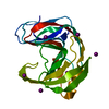
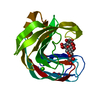
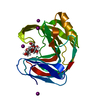
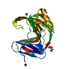
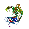
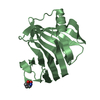
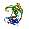
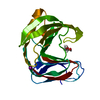
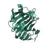
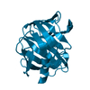
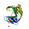
- PDB-6jxl: Crystal Structures of Endo-beta-1,4-xylanase II Complexed with Xy... -
+ Open data

- Basic information
| Entry | Database: PDB / ID: 6jxl | |||||||||
|---|---|---|---|---|---|---|---|---|---|---|
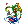
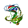
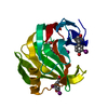
| Title | Crystal Structures of Endo-beta-1,4-xylanase II Complexed with Xylotriose | |||||||||
 Components Components | Endo-1,4-beta-xylanase 2 | |||||||||
 Keywords Keywords | HYDROLASE / xylanase II / complex / Xylotriose | |||||||||
| Function / homology |  Function and homology information Function and homology informationendo-1,4-beta-xylanase / endo-1,4-beta-xylanase activity / xylan catabolic process / extracellular region Similarity search - Function | |||||||||
| Biological species |  Trichoderma reesei RUT C-30 (fungus) Trichoderma reesei RUT C-30 (fungus) | |||||||||
| Method |  X-RAY DIFFRACTION / SYNCHROTRON / MOLECULAR REPLACEMENT / Resolution: 1.3 Å X-RAY DIFFRACTION / SYNCHROTRON / MOLECULAR REPLACEMENT / Resolution: 1.3 Å | |||||||||
 Authors Authors | Li, Z. / Wan, Q. | |||||||||
| Funding support |  China, 1items China, 1items
| |||||||||
 Citation Citation | Journal: To Be Published Title: Crystal Structures of Endo-beta-1,4-xylanase II Complexed with Xylotriose Authors: Wan, Q. #1: Journal: ACTA CRYSTALLOGR.,SECT.D / Year: 2014Title: X-ray crystallographic studies of family 11 xylanase Michaelis and product complexes: implications for the catalytic mechanism Authors: Wan, Q. / Qiu, Z. #2: Journal: PROC.NATL.ACAD.SCI.USA / Year: 2015Title: Direct determination of protonation states and visualization of hydrogen bonding in a glycoside hydrolase with neutron crystallography Authors: Wan, Q. / Jerry, M.P. | |||||||||
| History |
|
- Structure visualization
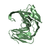
| Structure viewer | Molecule: MolmilJmol/JSmol |
|---|
- Downloads & links
-Download
| PDBx/mmCIF format | 6jxl.cif.gz | 127.4 KB | Display | PDBx/mmCIF format |
|---|---|---|---|---|
| PDB format | pdb6jxl.ent.gz | 99.9 KB | Display | PDB format |
| PDBx/mmJSON format | 6jxl.json.gz | Tree view | PDBx/mmJSON format | |
| Others |  Other downloads Other downloads |
-Validation report
| Arichive directory | https://data.pdbj.org/pub/pdb/validation_reports/jx/6jxlftp://data.pdbj.org/pub/pdb/validation_reports/jx/6jxl | HTTPS FTP |
|---|
-Related structure data
| Related structure data |  5zf3C  2dfcS S: Starting model for refinement C: citing same article ( |
|---|---|
| Similar structure data | |
| Experimental dataset #1 | Data reference: 10.1073/pnas.1504986112 / Data set type: diffraction image data |


-Links
 PDBj
PDBj
- Assembly
| Deposited unit | 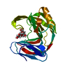
| ||||||||
|---|---|---|---|---|---|---|---|---|---|
| 1 |
| ||||||||
| Unit cell |
|
-Components
| #1: Protein | Mass: 20728.322 Da / Num. of mol.: 1 / Fragment: UNP residues 35-223 / Mutation: N44D Source method: isolated from a genetically manipulated source Source: (gene. exp.) Trichoderma reesei RUT C-30 (fungus) / Strain: Rut C-30 / Gene: xyn2 / Production host:  |
|---|---|
| #2: Polysaccharide | beta-D-xylopyranose-(1-4)-beta-D-xylopyranose-(1-4)-beta-D-xylopyranose / 4beta-beta-xylotriose  Source method: isolated from a genetically manipulated source Details: oligosaccharide / References: 4beta-beta-xylotriose |
| #3: Chemical | ChemComp-IOD /   Mass: 126.904 Da / Num. of mol.: 1 / Source method: obtained synthetically / Formula: I Mass: 126.904 Da / Num. of mol.: 1 / Source method: obtained synthetically / Formula: I |
| #4: Water | ChemComp-HOH /  Mass: 18.015 Da / Num. of mol.: 234 / Source method: isolated from a natural source / Formula: H2O Mass: 18.015 Da / Num. of mol.: 234 / Source method: isolated from a natural source / Formula: H2O |
| Has protein modification | N |
-Experimental details
-Experiment
| Experiment | Method: X-RAY DIFFRACTION / Number of used crystals: 1 |
|---|
- Sample preparation
| Crystal | Density Matthews: 2.35 Å3/Da / Density % sol: 47.66 % |
|---|---|
| Crystal grow | Temperature: 280 K / Method: evaporation / pH: 6 / Details: 20%PEG 8000, 0.2M NaI,0.1M MES |
-Data collection
| Diffraction | Mean temperature: 280 K / Serial crystal experiment: N |
|---|---|
| Diffraction source | Source: SYNCHROTRON / Site: SSRF / Beamline: BL19U1 / Wavelength: 1 Å |
| Detector | Type: ADSC QUANTUM 315r / Detector: CCD / Date: Nov 7, 2018 |
| Radiation | Protocol: SINGLE WAVELENGTH / Monochromatic (M) / Laue (L): M / Scattering type: x-ray |
| Radiation wavelength | Wavelength: 1 Å / Relative weight: 1 |
| Reflection | Resolution: 1.3→44.965 Å / Num. obs: 39057 / % possible obs: 80.04 % / Redundancy: 5.5 % / Biso Wilson estimate: 13.4 Å2 / Net I/σ(I): 14.7 |
| Reflection shell | Resolution: 1.3→1.347 Å / Mean I/σ(I) obs: 1.7 / Num. unique obs: 1793 |
- Processing
| Software |
| |||||||||||||||||||||||||||||||||||||||||||||||||||||||||||||||||||||||||||||||||||||||||||||||||||||||||
|---|---|---|---|---|---|---|---|---|---|---|---|---|---|---|---|---|---|---|---|---|---|---|---|---|---|---|---|---|---|---|---|---|---|---|---|---|---|---|---|---|---|---|---|---|---|---|---|---|---|---|---|---|---|---|---|---|---|---|---|---|---|---|---|---|---|---|---|---|---|---|---|---|---|---|---|---|---|---|---|---|---|---|---|---|---|---|---|---|---|---|---|---|---|---|---|---|---|---|---|---|---|---|---|---|---|---|
| Refinement | Method to determine structure: MOLECULAR REPLACEMENT Starting model: 2DFC Resolution: 1.3→32.67 Å / SU ML: 0.12 / Cross valid method: THROUGHOUT / σ(F): 1.34 / Phase error: 19.29
| |||||||||||||||||||||||||||||||||||||||||||||||||||||||||||||||||||||||||||||||||||||||||||||||||||||||||
| Solvent computation | Shrinkage radii: 0.9 Å / VDW probe radii: 1.11 Å | |||||||||||||||||||||||||||||||||||||||||||||||||||||||||||||||||||||||||||||||||||||||||||||||||||||||||
| Displacement parameters | Biso max: 50.77 Å2 / Biso mean: 18.7787 Å2 / Biso min: 7.99 Å2 | |||||||||||||||||||||||||||||||||||||||||||||||||||||||||||||||||||||||||||||||||||||||||||||||||||||||||
| Refinement step | Cycle: final / Resolution: 1.3→32.67 Å
| |||||||||||||||||||||||||||||||||||||||||||||||||||||||||||||||||||||||||||||||||||||||||||||||||||||||||
| Refine LS restraints |
| |||||||||||||||||||||||||||||||||||||||||||||||||||||||||||||||||||||||||||||||||||||||||||||||||||||||||
| LS refinement shell | Refine-ID: X-RAY DIFFRACTION / Rfactor Rfree error: 0 / Total num. of bins used: 14
|
