 Movie
Movie Controller


+ Open data

- Basic information
| Entry | Database: PDB / ID: 6jpf | ||||||||||||
|---|---|---|---|---|---|---|---|---|---|---|---|---|---|
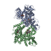
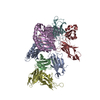
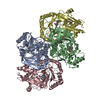
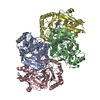
| Title | Structure of atOSCA1.1 channel at 3.52A | ||||||||||||
 Components Components | Protein OSCA1 | ||||||||||||
 Keywords Keywords | METAL TRANSPORT / osca / TMEM63 / ion channel / mechanosensitive / membrane protein / osmosensing | ||||||||||||
| Function / homology |  Function and homology information Function and homology informationregulation of calcium ion import / mechanosensitive monoatomic ion channel activity / cellular hyperosmotic response / calcium-activated cation channel activity / response to osmotic stress / monoatomic cation channel activity / protein tetramerization / plasma membrane / cytosol Similarity search - Function | ||||||||||||
| Biological species |  | ||||||||||||
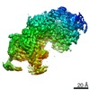
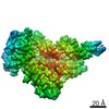
| Method | ELECTRON MICROSCOPY / single particle reconstruction / cryo EM / Resolution: 3.52 Å | ||||||||||||
 Authors Authors | Chen, L. / Zhang, M. / Kang, Y. / Wu, J.X. | ||||||||||||
| Funding support |  China, 3items China, 3items
| ||||||||||||
 Citation Citation | Journal: Nat Struct Mol Biol / Year: 2018 Title: Structure of the mechanosensitive OSCA channels. Authors: Mingfeng Zhang / Dali Wang / Yunlu Kang / Jing-Xiang Wu / Fuqiang Yao / Chengfang Pan / Zhiqiang Yan / Chen Song / Lei Chen / Abstract: Mechanosensitive ion channels convert mechanical stimuli into a flow of ions. These channels are widely distributed from bacteria to higher plants and humans, and are involved in many crucial ...Mechanosensitive ion channels convert mechanical stimuli into a flow of ions. These channels are widely distributed from bacteria to higher plants and humans, and are involved in many crucial physiological processes. Here we show that two members of the OSCA protein family in Arabidopsis thaliana, namely AtOSCA1.1 and AtOSCA3.1, belong to a new class of mechanosensitive ion channels. We solve the structure of the AtOSCA1.1 channel at 3.5-Å resolution and AtOSCA3.1 at 4.8-Å resolution by cryo-electron microscopy. OSCA channels are symmetric dimers that are mediated by cytosolic inter-subunit interactions. Strikingly, they have structural similarity to the mammalian TMEM16 family proteins. Our structural analysis accompanied with electrophysiological studies identifies the ion permeation pathway within each subunit and suggests a conformational change model for activation. | ||||||||||||
| History |
|
- Structure visualization
| Movie |
Movie viewer |
|---|---|
| Structure viewer | Molecule: MolmilJmol/JSmol |
- Downloads & links
-Download
| PDBx/mmCIF format | 6jpf.cif.gz | 245.7 KB | Display | PDBx/mmCIF format |
|---|---|---|---|---|
| PDB format | pdb6jpf.ent.gz | 196.4 KB | Display | PDB format |
| PDBx/mmJSON format | 6jpf.json.gz | Tree view | PDBx/mmJSON format | |
| Others |  Other downloads Other downloads |
-Validation report
| Arichive directory | https://data.pdbj.org/pub/pdb/validation_reports/jp/6jpfftp://data.pdbj.org/pub/pdb/validation_reports/jp/6jpf | HTTPS FTP |
|---|
-Related structure data
| Related structure data |  6822MC  6875C  5z1fC M: map data used to model this data C: citing same article ( |
|---|---|
| Similar structure data |


-Links
 PDBj
PDBj- Assembly
| Deposited unit | 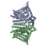
|
|---|---|
| 1 |
|
-Components
| #1: Protein | Mass: 87697.008 Da / Num. of mol.: 2 Source method: isolated from a genetically manipulated source Source: (gene. exp.)  Homo sapiens (human) / References: UniProt: Q9XEA1 Homo sapiens (human) / References: UniProt: Q9XEA1 |
|---|
-Experimental details
-Experiment
| Experiment | Method: ELECTRON MICROSCOPY |
|---|---|
| EM experiment | Aggregation state: PARTICLE / 3D reconstruction method: single particle reconstruction |
- Sample preparation
| Component | Name: atOSCA1.1 channel in detergent / Type: COMPLEX / Entity ID: all / Source: RECOMBINANT |
|---|---|
| Source (natural) | Organism: |
| Source (recombinant) | Organism: Homo sapiens (human) |
| Buffer solution | pH: 8 |
| Specimen | Embedding applied: NO / Shadowing applied: NO / Staining applied: NO / Vitrification applied: YES |
| Vitrification | Cryogen name: ETHANE |
- Electron microscopy imaging
| Experimental equipment |  Model: Titan Krios / Image courtesy: FEI Company |
|---|---|
| Microscopy | Model: FEI TITAN KRIOS |
| Electron gun | Electron source:  FIELD EMISSION GUN / Accelerating voltage: 300 kV / Illumination mode: FLOOD BEAM FIELD EMISSION GUN / Accelerating voltage: 300 kV / Illumination mode: FLOOD BEAM |
| Electron lens | Mode: BRIGHT FIELD |
| Image recording | Electron dose: 50 e/Å2 / Film or detector model: GATAN K2 SUMMIT (4k x 4k) |
- Processing
| CTF correction | Type: NONE |
|---|---|
| 3D reconstruction | Resolution: 3.52 Å / Resolution method: FSC 0.143 CUT-OFF / Num. of particles: 115697 / Symmetry type: POINT |
