 Movie
Movie Controller

[English] 日本語
 Yorodumi
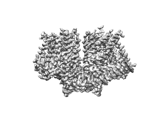
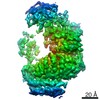
- EMDB-9112: Cryo-EM map of mechanically activated ion channel OSCA1.2 in nanodisc -
+ Open data

- Basic information
| Entry | Database: EMDB / ID: EMD-9112 | |||||||||
|---|---|---|---|---|---|---|---|---|---|---|
| Title | Cryo-EM map of mechanically activated ion channel OSCA1.2 in nanodisc | |||||||||
 Map data Map data | Cryo-EM map of mechanically activated ion channel OSCA1.2 in nanodisc | |||||||||
 Sample Sample |
| |||||||||
 Keywords Keywords | Mechanically activated ion channel / MEMBRANE PROTEIN | |||||||||
| Function / homology |  Function and homology information Function and homology informationmechanosensitive monoatomic ion channel activity / calcium-activated cation channel activity / monoatomic cation transport / identical protein binding / plasma membrane Similarity search - Function | |||||||||
| Biological species |  | |||||||||
| Method | single particle reconstruction / cryo EM / Resolution: 3.1 Å | |||||||||
 Authors Authors | Jojoa-Cruz S / Saotome K | |||||||||
| Funding support |  United States, 2 items United States, 2 items
| |||||||||
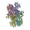
 Citation Citation | Journal: Elife / Year: 2018 Title: Cryo-EM structure of the mechanically activated ion channel OSCA1.2. Authors: Sebastian Jojoa-Cruz / Kei Saotome / Swetha E Murthy / Che Chun Alex Tsui / Mark Sp Sansom / Ardem Patapoutian / Andrew B Ward /  Abstract: Mechanically activated ion channels underlie touch, hearing, shear-stress sensing, and response to turgor pressure. OSCA/TMEM63s are a newly-identified family of eukaryotic mechanically activated ion ...Mechanically activated ion channels underlie touch, hearing, shear-stress sensing, and response to turgor pressure. OSCA/TMEM63s are a newly-identified family of eukaryotic mechanically activated ion channels opened by membrane tension. The structural underpinnings of OSCA/TMEM63 function are not explored. Here, we elucidate high resolution cryo-electron microscopy structures of OSCA1.2, revealing a dimeric architecture containing eleven transmembrane helices per subunit and surprising topological similarities to TMEM16 proteins. We locate the ion permeation pathway within each subunit by demonstrating that a conserved acidic residue is a determinant of channel conductance. Molecular dynamics simulations reveal membrane interactions, suggesting the role of lipids in OSCA1.2 gating. These results lay a foundation to decipher how the structural organization of OSCA/TMEM63 is suited for their roles as MA ion channels. | |||||||||
| History |
|
- Structure visualization
| Movie |
Movie viewer |
|---|---|
| Structure viewer | EM map: SurfViewMolmilJmol/JSmol |
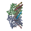
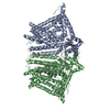
| Supplemental images |
- Downloads & links
-EMDB archive
| Map data | emd_9112.map.gz | 28.5 MB | EMDB map data format | |
|---|---|---|---|---|
| Header (meta data) | emd-9112-v30.xmlemd-9112.xml | 16.7 KB 16.7 KB | Display Display | EMDB header |
| FSC (resolution estimation) | emd_9112_fsc.xml | 7.1 KB | Display | FSC data file |
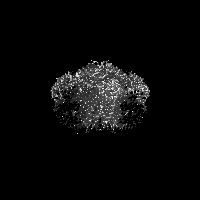
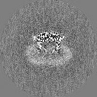
| Images |  emd_9112.png emd_9112.png | 74.8 KB | ||
| Filedesc metadata | emd-9112.cif.gz | 6.1 KB | ||
| Others | emd_9112_half_map_1.map.gzemd_9112_half_map_2.map.gz | 22.4 MB 22.4 MB | ||
| Archive directory |  http://ftp.pdbj.org/pub/emdb/structures/EMD-9112ftp://ftp.pdbj.org/pub/emdb/structures/EMD-9112 http://ftp.pdbj.org/pub/emdb/structures/EMD-9112ftp://ftp.pdbj.org/pub/emdb/structures/EMD-9112 | HTTPS FTP |
-Related structure data
| Related structure data |  6mgvMC  9113C  6mgwC C: citing same article ( M: atomic model generated by this map |
|---|---|
| Similar structure data |


-Links
| EMDB pages | EMDB (EBI/PDBe) / EMDataResource |
|---|
-Map
| File | Download / File: emd_9112.map.gz / Format: CCP4 / Size: 30.5 MB / Type: IMAGE STORED AS FLOATING POINT NUMBER (4 BYTES) | ||||||||||||||||||||||||||||||||||||||||||||||||||||||||||||
|---|---|---|---|---|---|---|---|---|---|---|---|---|---|---|---|---|---|---|---|---|---|---|---|---|---|---|---|---|---|---|---|---|---|---|---|---|---|---|---|---|---|---|---|---|---|---|---|---|---|---|---|---|---|---|---|---|---|---|---|---|---|
| Annotation | Cryo-EM map of mechanically activated ion channel OSCA1.2 in nanodisc | ||||||||||||||||||||||||||||||||||||||||||||||||||||||||||||
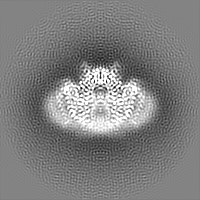
| Projections & slices | Image control
Images are generated by Spider. | ||||||||||||||||||||||||||||||||||||||||||||||||||||||||||||
| Voxel size | X=Y=Z: 1.15 Å | ||||||||||||||||||||||||||||||||||||||||||||||||||||||||||||
| Density |
| ||||||||||||||||||||||||||||||||||||||||||||||||||||||||||||
| Symmetry | Space group: 1 | ||||||||||||||||||||||||||||||||||||||||||||||||||||||||||||
| Details | EMDB XML:
CCP4 map header:
| ||||||||||||||||||||||||||||||||||||||||||||||||||||||||||||
 Z (Sec.)
 Y (Row.)
 X (Col.)


-Supplemental data
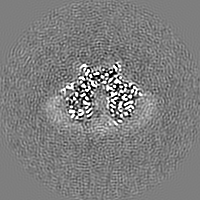
-Half map: Half map 1
| File | emd_9112_half_map_1.map | ||||||||||||
|---|---|---|---|---|---|---|---|---|---|---|---|---|---|
| Annotation | Half map 1 | ||||||||||||
| Projections & Slices |
| ||||||||||||
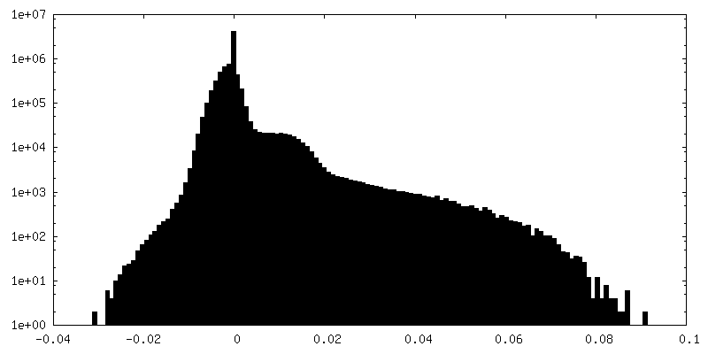
| Density Histograms |


-Half map: Half map 2
| File | emd_9112_half_map_2.map | ||||||||||||
|---|---|---|---|---|---|---|---|---|---|---|---|---|---|
| Annotation | Half map 2 | ||||||||||||
| Projections & Slices |
| ||||||||||||
| Density Histograms |


- Sample components
-Entire : OSCA1.2
| Entire | Name: OSCA1.2 |
|---|---|
| Components |
|
-Supramolecule #1: OSCA1.2
| Supramolecule | Name: OSCA1.2 / type: organelle_or_cellular_component / ID: 1 / Parent: 0 / Macromolecule list: all |
|---|---|
| Source (natural) | Organism: |
| Molecular weight | Theoretical: 88 KDa |
-Macromolecule #1: Calcium permeable stress-gated cation channel 1
| Macromolecule | Name: Calcium permeable stress-gated cation channel 1 / type: protein_or_peptide / ID: 1 / Number of copies: 2 / Enantiomer: LEVO |
|---|---|
| Source (natural) | Organism: |
| Molecular weight | Theoretical: 89.031719 KDa |
| Recombinant expression | Organism:  Homo sapiens (human) Homo sapiens (human) |
| Sequence | String: MATLQDIGVS AGINILSAFV FFIIFAVLRL QPFNDRVYFS KWYLKGLRSS PARGGAFAQR FVNLDFRSYM KFLNWMPEAL KMPEPELID HAGLDSVVYL RIYWLGLKIF TPIAVLAWAV LVPVNWTNNT LEMAKQLRNV TSSDIDKLSV SNIPEYSMRF W THIVMAYA ...String: MATLQDIGVS AGINILSAFV FFIIFAVLRL QPFNDRVYFS KWYLKGLRSS PARGGAFAQR FVNLDFRSYM KFLNWMPEAL KMPEPELID HAGLDSVVYL RIYWLGLKIF TPIAVLAWAV LVPVNWTNNT LEMAKQLRNV TSSDIDKLSV SNIPEYSMRF W THIVMAYA FTIWTCYVLM KEYETIANMR LQFVASEARR PDQFTVLVRN VPPDADESVS ELVEHFFLVN HPDHYLTHQV VC NANKLAD LVKKKKKLQN WLDYYQLKYA RNNSQRIMVK LGFLGLWGQK VDAIEHYIAE IDKISKEISK EREEVVNDPK AIM PAAFVS FKTRWAAAVC AQTQQTRNPT QWLTEWAPEP RDVFWSNLAI PYVSLTVRRL IMHVAFFFLT FFFIVPIAFV QSLA TIEGI VKAAPFLKFI VDDKFMKSVI QGFLPGIALK LFLAFLPSIL MIMSKFEGFT SISSLERRAA FRYYIFNLVN VFLAS VIAG AAFEQLNSFL NQSANQIPKT IGVAIPMKAT FFITYIMVDG WAGVAGEILM LKPLIMFHLK NAFLVKTDKD REEAMD PGS IGFNTGEPRI QLYFLLGLVY APVTPMLLPF ILVFFALAYI VYRHQIINVY NQEYESAAAF WPDVHGRVIA ALVISQL LL MGLLGTKHAA LAAPFLIALP VLTIGFHHFC KGRYEPAFIR YPLQEAMMKD TLETAREPNL NLKGYLQNAY VHPVFKGD E DDYDIDDKLG KFEDEAIIVP TKRQSRRNTP APSIISGDDS PSLPFSGKLV GTGTLEVLFQ UniProtKB: Hyperosmolality-gated Ca2+ permeable channel 1.2 |
-Experimental details
-Structure determination
| Method | cryo EM |
|---|---|
 Processing Processing | single particle reconstruction |
| Aggregation state | particle |
-Sample preparation
| Concentration | 2.1 mg/mL |
|---|---|
| Buffer | pH: 8 |
| Grid | Material: GOLD / Mesh: 300 |
| Vitrification | Cryogen name: ETHANE / Instrument: FEI VITROBOT MARK IV |
- Electron microscopy
| Microscope | FEI TALOS ARCTICA |
|---|---|
| Image recording | Film or detector model: GATAN K2 SUMMIT (4k x 4k) / Detector mode: COUNTING / Digitization - Dimensions - Width: 3710 pixel / Digitization - Dimensions - Height: 3838 pixel / Number grids imaged: 1 / Number real images: 1740 / Average electron dose: 60.0 e/Å2 |
| Electron beam | Acceleration voltage: 200 kV / Electron source:  FIELD EMISSION GUN FIELD EMISSION GUN |
| Electron optics | C2 aperture diameter: 70.0 µm / Illumination mode: OTHER / Imaging mode: BRIGHT FIELD / Cs: 2.7 mm / Nominal defocus max: -2.2 µm / Nominal defocus min: -0.4 µm / Nominal magnification: 36000 |
| Experimental equipment |  Model: Talos Arctica / Image courtesy: FEI Company |

