 Movie
Movie Controller

[English] 日本語
 Yorodumi
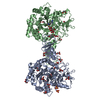
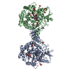
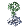
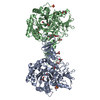
- PDB-6jkf: Crystal structure of Serratia marcescens Chitinase B complexed wi... -
+ Open data

- Basic information
| Entry | Database: PDB / ID: 6jkf | ||||||
|---|---|---|---|---|---|---|---|
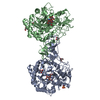
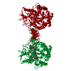
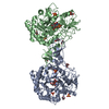
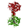
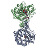
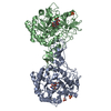
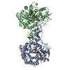
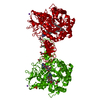
| Title | Crystal structure of Serratia marcescens Chitinase B complexed with compound 2-8-s2 | ||||||
 Components Components | Chitinase | ||||||
 Keywords Keywords | HYDROLASE / chitinase / complex / Serratia marcescens | ||||||
| Function / homology |  Function and homology information Function and homology informationendochitinase activity / chitinase / chitin catabolic process / chitin binding / polysaccharide catabolic process / carbohydrate binding / extracellular region Similarity search - Function | ||||||
| Biological species |  Serratia marcescens (bacteria) Serratia marcescens (bacteria) | ||||||
| Method |  X-RAY DIFFRACTION / SYNCHROTRON / MOLECULAR REPLACEMENT / Resolution: 1.99 Å X-RAY DIFFRACTION / SYNCHROTRON / MOLECULAR REPLACEMENT / Resolution: 1.99 Å | ||||||
 Authors Authors | Yang, Q. / Jiang, X. | ||||||
| Funding support |  China, 1items China, 1items
| ||||||
 Citation Citation | Journal: J.Med.Chem. / Year: 2020 Title: A Series of Compounds Bearing a Dipyrido-Pyrimidine Scaffold Acting as Novel Human and Insect Pest Chitinase Inhibitors. Authors: Jiang, X. / Kumar, A. / Motomura, Y. / Liu, T. / Zhou, Y. / Moro, K. / Zhang, K.Y.J. / Yang, Q. | ||||||
| History |
|
- Structure visualization
| Structure viewer | Molecule: MolmilJmol/JSmol |
|---|
- Downloads & links
-Download
| PDBx/mmCIF format | 6jkf.cif.gz | 222.3 KB | Display | PDBx/mmCIF format |
|---|---|---|---|---|
| PDB format | pdb6jkf.ent.gz | 174.8 KB | Display | PDB format |
| PDBx/mmJSON format | 6jkf.json.gz | Tree view | PDBx/mmJSON format | |
| Others |  Other downloads Other downloads |
-Validation report
| Arichive directory | https://data.pdbj.org/pub/pdb/validation_reports/jk/6jkfftp://data.pdbj.org/pub/pdb/validation_reports/jk/6jkf | HTTPS FTP |
|---|
-Related structure data
| Related structure data |  6jjrC 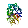 6jk6C  6jk9C 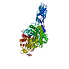 6jmnC  4z2gS S: Starting model for refinement C: citing same article ( |
|---|---|
| Similar structure data |


-Links
 PDBj
PDBj- Assembly
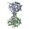
| Deposited unit | 
| ||||||||
|---|---|---|---|---|---|---|---|---|---|
| 1 |
| ||||||||
| Unit cell |
|
-Components
| #1: Protein | Mass: 55299.742 Da / Num. of mol.: 2 Source method: isolated from a genetically manipulated source Source: (gene. exp.) Serratia marcescens (bacteria) / Gene: chiB / Plasmid: pet28a / Production host: #2: Chemical | 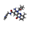  Mass: 445.494 Da / Num. of mol.: 3 / Source method: obtained synthetically / Formula: C24H25N6O3 Mass: 445.494 Da / Num. of mol.: 3 / Source method: obtained synthetically / Formula: C24H25N6O3#3: Water | ChemComp-HOH / |  Mass: 18.015 Da / Num. of mol.: 691 / Source method: isolated from a natural source / Formula: H2O Mass: 18.015 Da / Num. of mol.: 691 / Source method: isolated from a natural source / Formula: H2OHas ligand of interest | Y | Has protein modification | Y | |
|---|
-Experimental details
-Experiment
| Experiment | Method: X-RAY DIFFRACTION / Number of used crystals: 1 |
|---|
- Sample preparation
| Crystal | Density Matthews: 2.46 Å3/Da / Density % sol: 50.03 % |
|---|---|
| Crystal grow | Temperature: 293 K / Method: vapor diffusion, hanging drop / pH: 5.6 Details: 50 mM citrate (pH 5.6), 0.5 M Li2SO4, and 0.25 M (NH4)2SO4 |
-Data collection
| Diffraction | Mean temperature: 100 K / Serial crystal experiment: N |
|---|---|
| Diffraction source | Source: SYNCHROTRON / Site: NFPSS / Beamline: BL18U / Wavelength: 0.97778 Å |
| Detector | Type: ADSC QUANTUM 315r / Detector: CCD / Date: Jan 20, 2016 |
| Radiation | Protocol: SINGLE WAVELENGTH / Monochromatic (M) / Laue (L): M / Scattering type: x-ray |
| Radiation wavelength | Wavelength: 0.97778 Å / Relative weight: 1 |
| Reflection | Resolution: 1.99→29.712 Å / Num. obs: 75549 / % possible obs: 100 % / Redundancy: 13.1 % / Rsym value: 0.109 / Net I/σ(I): 25.5 |
| Reflection shell | Resolution: 2→2.03 Å / Redundancy: 13.3 % / Mean I/σ(I) obs: 4.4 / Num. unique obs: 3683 / Rsym value: 0.375 / % possible all: 100 |
- Processing
| Software |
| |||||||||||||||||||||||||||||||||||||||||||||||||||||||||||||||||||||||||||||||||||||||||||||||||||||||||
|---|---|---|---|---|---|---|---|---|---|---|---|---|---|---|---|---|---|---|---|---|---|---|---|---|---|---|---|---|---|---|---|---|---|---|---|---|---|---|---|---|---|---|---|---|---|---|---|---|---|---|---|---|---|---|---|---|---|---|---|---|---|---|---|---|---|---|---|---|---|---|---|---|---|---|---|---|---|---|---|---|---|---|---|---|---|---|---|---|---|---|---|---|---|---|---|---|---|---|---|---|---|---|---|---|---|---|
| Refinement | Method to determine structure: MOLECULAR REPLACEMENT Starting model: 4Z2G Resolution: 1.99→29.712 Å / SU ML: 0.15 / Cross valid method: FREE R-VALUE / σ(F): 1.34 / Phase error: 20.85
| |||||||||||||||||||||||||||||||||||||||||||||||||||||||||||||||||||||||||||||||||||||||||||||||||||||||||
| Solvent computation | Shrinkage radii: 0.9 Å / VDW probe radii: 1.11 Å | |||||||||||||||||||||||||||||||||||||||||||||||||||||||||||||||||||||||||||||||||||||||||||||||||||||||||
| Refinement step | Cycle: LAST / Resolution: 1.99→29.712 Å
| |||||||||||||||||||||||||||||||||||||||||||||||||||||||||||||||||||||||||||||||||||||||||||||||||||||||||
| Refine LS restraints |
| |||||||||||||||||||||||||||||||||||||||||||||||||||||||||||||||||||||||||||||||||||||||||||||||||||||||||
| LS refinement shell |
|
