 Movie
Movie Controller

[English] 日本語
 Yorodumi
- PDB-1w1v: Crystal structure of S. marcescens chitinase B in complex with th... -
+ Open data

- Basic information
| Entry | Database: PDB / ID: 1w1v | ||||||
|---|---|---|---|---|---|---|---|
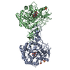
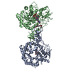
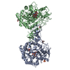
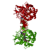
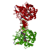
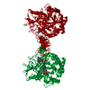
| Title | Crystal structure of S. marcescens chitinase B in complex with the cyclic dipeptide inhibitor cyclo-(L-Arg-L-Pro) at 1.85 A resolution | ||||||
 Components Components | CHITINASE B | ||||||
 Keywords Keywords | HYDROLASE / GLYCOSIDE HYDROLASE / CHITINASE / STRUCTURE-BASED INHIBITOR DESIGN / CYCLIC DIPEPTIDE | ||||||
| Function / homology |  Function and homology information Function and homology informationendochitinase activity / chitinase / chitin catabolic process / chitin binding / polysaccharide catabolic process / carbohydrate binding / extracellular region Similarity search - Function | ||||||
| Biological species |  SERRATIA MARCESCENS (bacteria) SERRATIA MARCESCENS (bacteria) | ||||||
| Method |  X-RAY DIFFRACTION / MOLECULAR REPLACEMENT / Resolution: 1.85 Å X-RAY DIFFRACTION / MOLECULAR REPLACEMENT / Resolution: 1.85 Å | ||||||
 Authors Authors | Houston, D.R. / Synstad, B. / Eijsink, V.G.H. / Eggleston, I. / Van Aalten, D.M.F. | ||||||
 Citation Citation | Journal: J.Med.Chem. / Year: 2004 Title: Structure-Based Exploration of Cyclic Dipeptide Chitinase Inhibitors Authors: Houston, D.R. / Synstad, B. / Eijsink, V.G.H. / Stark, M.J. / Eggleston, I. / Van Aalten, D.M.F. | ||||||
| History |
| ||||||
| Remark 700 | SHEET DETERMINATION METHOD: DSSP THE SHEETS PRESENTED AS "AA" IN EACH CHAIN ON SHEET RECORDS BELOW ... SHEET DETERMINATION METHOD: DSSP THE SHEETS PRESENTED AS "AA" IN EACH CHAIN ON SHEET RECORDS BELOW IS ACTUALLY AN 10-STRANDED BARREL THIS IS REPRESENTED BY A 11-STRANDED SHEET IN WHICH THE FIRST AND LAST STRANDS ARE IDENTICAL. THE SHEETS PRESENTED AS "BA" IN EACH CHAIN ON SHEET RECORDS BELOW IS ACTUALLY AN 9-STRANDED BARREL THIS IS REPRESENTED BY A 10-STRANDED SHEET IN WHICH THE FIRST AND LAST STRANDS ARE IDENTICAL. |
- Structure visualization
| Structure viewer | Molecule: MolmilJmol/JSmol |
|---|
- Downloads & links
-Download
| PDBx/mmCIF format | 1w1v.cif.gz | 224.8 KB | Display | PDBx/mmCIF format |
|---|---|---|---|---|
| PDB format | pdb1w1v.ent.gz | 181.8 KB | Display | PDB format |
| PDBx/mmJSON format | 1w1v.json.gz | Tree view | PDBx/mmJSON format | |
| Others |  Other downloads Other downloads |
-Validation report
| Arichive directory | https://data.pdbj.org/pub/pdb/validation_reports/w1/1w1vftp://data.pdbj.org/pub/pdb/validation_reports/w1/1w1v | HTTPS FTP |
|---|
-Related structure data
| Related structure data |  1w1pC  1w1tC  1w1yC  1o6iS C: citing same article ( S: Starting model for refinement |
|---|---|
| Similar structure data |


-Links
 PDBj
PDBj- Assembly
| Deposited unit | 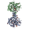
| ||||||||
|---|---|---|---|---|---|---|---|---|---|
| 1 |
| ||||||||
| Unit cell |
|
-Components
| #1: Protein | Mass: 55518.012 Da / Num. of mol.: 2 Source method: isolated from a genetically manipulated source Source: (gene. exp.) SERRATIA MARCESCENS (bacteria) / Production host: #2: Chemical | ChemComp-GOL /   Mass: 92.094 Da / Num. of mol.: 31 / Source method: obtained synthetically / Formula: C3H8O3 Mass: 92.094 Da / Num. of mol.: 31 / Source method: obtained synthetically / Formula: C3H8O3#3: Chemical |   Mass: 96.063 Da / Num. of mol.: 2 / Source method: obtained synthetically / Formula: SO4 Mass: 96.063 Da / Num. of mol.: 2 / Source method: obtained synthetically / Formula: SO4#4: Chemical |   Mass: 253.301 Da / Num. of mol.: 2 / Source method: obtained synthetically / Formula: C11H19N5O2 Mass: 253.301 Da / Num. of mol.: 2 / Source method: obtained synthetically / Formula: C11H19N5O2#5: Water | ChemComp-HOH / |  Mass: 18.015 Da / Num. of mol.: 707 / Source method: isolated from a natural source / Formula: H2O Mass: 18.015 Da / Num. of mol.: 707 / Source method: isolated from a natural source / Formula: H2OHas protein modification | Y | |
|---|
-Experimental details
-Experiment
| Experiment | Method: X-RAY DIFFRACTION / Number of used crystals: 1 |
|---|
- Sample preparation
| Crystal | Density Matthews: 2.43 Å3/Da / Density % sol: 48 % |
|---|---|
| Crystal grow | pH: 7 Details: 1.5 M AMMONIUM SULPHATE, 0.1 M HEPES PH 7, 25 % GLYCEROL |
-Data collection
| Diffraction | Mean temperature: 100 K |
|---|---|
| Diffraction source | Source: ROTATING ANODE / Type: RIGAKU / Wavelength: 1.54 |
| Detector | Type: RIGAKU RAXIS-IV / Detector: IMAGE PLATE / Details: MIRRORS |
| Radiation | Protocol: SINGLE WAVELENGTH / Monochromatic (M) / Laue (L): M / Scattering type: x-ray |
| Radiation wavelength | Wavelength: 1.54 Å / Relative weight: 1 |
| Reflection | Resolution: 1.85→25 Å / Num. obs: 88471 / % possible obs: 95.5 % / Observed criterion σ(I): 0 / Redundancy: 3.11 % / Biso Wilson estimate: 18.5 Å2 / Rmerge(I) obs: 0.05 / Net I/σ(I): 30.1 |
| Reflection shell | Resolution: 1.85→1.92 Å / Rmerge(I) obs: 0.18 / Mean I/σ(I) obs: 6.96 / % possible all: 76.1 |
- Processing
| Software |
| ||||||||||||||||||||||||||||||||||||||||||||||||||||||||||||||||||||||||||||||||
|---|---|---|---|---|---|---|---|---|---|---|---|---|---|---|---|---|---|---|---|---|---|---|---|---|---|---|---|---|---|---|---|---|---|---|---|---|---|---|---|---|---|---|---|---|---|---|---|---|---|---|---|---|---|---|---|---|---|---|---|---|---|---|---|---|---|---|---|---|---|---|---|---|---|---|---|---|---|---|---|---|---|
| Refinement | Method to determine structure: MOLECULAR REPLACEMENT Starting model: PDB ENTRY 1O6I Resolution: 1.85→24.87 Å / Rfactor Rfree error: 0.007 / Data cutoff high absF: 2771894.21 / Cross valid method: THROUGHOUT / σ(F): 0 Stereochemistry target values: MAXIMUM LIKELIHOOD TARGET USING AMPLITUDES
| ||||||||||||||||||||||||||||||||||||||||||||||||||||||||||||||||||||||||||||||||
| Solvent computation | Solvent model: CNS BULK SOLVENT MODEL USED / Bsol: 43.853 Å2 / ksol: 0.374454 e/Å3 | ||||||||||||||||||||||||||||||||||||||||||||||||||||||||||||||||||||||||||||||||
| Displacement parameters | Biso mean: 28.47 Å2
| ||||||||||||||||||||||||||||||||||||||||||||||||||||||||||||||||||||||||||||||||
| Refine analyze |
| ||||||||||||||||||||||||||||||||||||||||||||||||||||||||||||||||||||||||||||||||
| Refinement step | Cycle: LAST / Resolution: 1.85→24.87 Å
| ||||||||||||||||||||||||||||||||||||||||||||||||||||||||||||||||||||||||||||||||
| Refine LS restraints |
| ||||||||||||||||||||||||||||||||||||||||||||||||||||||||||||||||||||||||||||||||
| LS refinement shell | Highest resolution: 1.85 Å |
