[English] 日本語
 Yorodumi
- PDB-6jig: Crystal structure of GMP reductase C318A from Trypanosoma brucei ... -
+ Open data
- Basic information
| Entry | Database: PDB / ID: 6jig | ||||||||||||
|---|---|---|---|---|---|---|---|---|---|---|---|---|---|
| Title | Crystal structure of GMP reductase C318A from Trypanosoma brucei in complex with guanosine 5'-monophosphate | ||||||||||||
 Components Components | GMP reductase | ||||||||||||
 Keywords Keywords | OXIDOREDUCTASE / Trypanosoma brucei / 5'-monophosphate reductase / guanosine 5'-monophosphate / cystathionine beta synthase motif | ||||||||||||
| Function / homology |  Function and homology information Function and homology informationGMP reductase activity / glycosome / IMP dehydrogenase activity / IMP dehydrogenase / GMP biosynthetic process / GTP biosynthetic process / GTP binding / nucleolus / metal ion binding / cytoplasm Similarity search - Function | ||||||||||||
| Biological species |  | ||||||||||||
| Method |  X-RAY DIFFRACTION / X-RAY DIFFRACTION /  SYNCHROTRON / SYNCHROTRON /  MOLECULAR REPLACEMENT / Resolution: 1.903 Å MOLECULAR REPLACEMENT / Resolution: 1.903 Å | ||||||||||||
 Authors Authors | Mase, H. / Imamura, A. / Nishimura, S. / Inui, T. | ||||||||||||
| Funding support |  Japan, 3items Japan, 3items
| ||||||||||||
 Citation Citation |  Journal: Nat Commun / Year: 2020 Journal: Nat Commun / Year: 2020Title: Allosteric regulation accompanied by oligomeric state changes of Trypanosoma brucei GMP reductase through cystathionine-beta-synthase domain. Authors: Imamura, A. / Okada, T. / Mase, H. / Otani, T. / Kobayashi, T. / Tamura, M. / Kubata, B.K. / Inoue, K. / Rambo, R.P. / Uchiyama, S. / Ishii, K. / Nishimura, S. / Inui, T. | ||||||||||||
| History |
|
- Structure visualization
| Structure viewer | Molecule:  Molmil Molmil Jmol/JSmol Jmol/JSmol |
|---|
- Downloads & links
- Download
| PDBx/mmCIF format |  6jig.cif.gz 6jig.cif.gz | 106.1 KB | Display |  PDBx/mmCIF format PDBx/mmCIF format |
|---|---|---|---|---|
| PDB format |  pdb6jig.ent.gz pdb6jig.ent.gz | 77.2 KB | Display |  PDB format PDB format |
| PDBx/mmJSON format |  6jig.json.gz 6jig.json.gz | Tree view |  PDBx/mmJSON format PDBx/mmJSON format | |
| Others |  Other downloads Other downloads |
-Validation report
| Arichive directory |  https://data.pdbj.org/pub/pdb/validation_reports/ji/6jig https://data.pdbj.org/pub/pdb/validation_reports/ji/6jig ftp://data.pdbj.org/pub/pdb/validation_reports/ji/6jig ftp://data.pdbj.org/pub/pdb/validation_reports/ji/6jig | HTTPS FTP |
|---|
-Related structure data
| Related structure data |  6jl8C  6lk4C  6ihy S: Starting model for refinement C: citing same article ( |
|---|---|
| Similar structure data |
- Links
Links
- Assembly
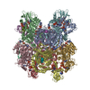
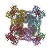
| Deposited unit | 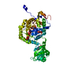
| ||||||||
|---|---|---|---|---|---|---|---|---|---|
| 1 | x 8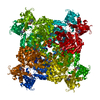
| ||||||||
| Unit cell |
|
- Components
| #1: Protein | Mass: 53806.180 Da / Num. of mol.: 1 / Mutation: C318A Source method: isolated from a genetically manipulated source Source: (gene. exp.)  Plasmid: pET22b / Production host:  | ||||||
|---|---|---|---|---|---|---|---|
| #2: Chemical | | #3: Chemical | ChemComp-K / | #4: Water | ChemComp-HOH / | Sequence details | The source organism of sequence reference UniProtKB entry Q57ZS7 (Q57ZS7_TRYB2), is described as ...The source organism of sequence reference UniProtKB entry Q57ZS7 (Q57ZS7_TRYB2), is described as 'Trypanosoma brucei brucei (strain 927/4 GUTat10.1)'. But, in this study, the gene for TbGMPR was isolated from Trypanosoma brucei brucei (strain ILTat1.4). The amino acid sequences of the enzymes from two strains are identical. | |
-Experimental details
-Experiment
| Experiment | Method:  X-RAY DIFFRACTION / Number of used crystals: 1 X-RAY DIFFRACTION / Number of used crystals: 1 |
|---|
- Sample preparation
| Crystal | Density Matthews: 3.17 Å3/Da / Density % sol: 61.2 % |
|---|---|
| Crystal grow | Temperature: 277 K / Method: vapor diffusion, sitting drop / pH: 7.5 / Details: 0.1M HEPES (pH 7.5), 0.75M NaH2PO4, 0.75M KH2PO4 |
-Data collection
| Diffraction | Mean temperature: 100 K / Serial crystal experiment: N |
|---|---|
| Diffraction source | Source:  SYNCHROTRON / Site: SYNCHROTRON / Site:  SPring-8 SPring-8  / Beamline: BL38B1 / Wavelength: 1 Å / Beamline: BL38B1 / Wavelength: 1 Å |
| Detector | Type: DECTRIS PILATUS3 6M / Detector: PIXEL / Date: Jul 4, 2018 |
| Radiation | Protocol: SINGLE WAVELENGTH / Monochromatic (M) / Laue (L): M / Scattering type: x-ray |
| Radiation wavelength | Wavelength: 1 Å / Relative weight: 1 |
| Reflection | Resolution: 1.9→48.73 Å / Num. obs: 54159 / % possible obs: 99.9 % / Redundancy: 25.5 % / CC1/2: 1 / Rmerge(I) obs: 0.039 / Net I/σ(I): 53.6 |
| Reflection shell | Resolution: 1.9→2.02 Å / Redundancy: 23.9 % / Rmerge(I) obs: 0.988 / Mean I/σ(I) obs: 3.6 / Num. unique obs: 8636 / CC1/2: 0.91 / % possible all: 99.5 |
- Processing
| Software |
| ||||||||||||||||||||||||||||||||||||||||||||||||||||||||||||||||||||||||||||||||||||||||||||||||||||||||||||||||||||||||||||||||||||||||||||
|---|---|---|---|---|---|---|---|---|---|---|---|---|---|---|---|---|---|---|---|---|---|---|---|---|---|---|---|---|---|---|---|---|---|---|---|---|---|---|---|---|---|---|---|---|---|---|---|---|---|---|---|---|---|---|---|---|---|---|---|---|---|---|---|---|---|---|---|---|---|---|---|---|---|---|---|---|---|---|---|---|---|---|---|---|---|---|---|---|---|---|---|---|---|---|---|---|---|---|---|---|---|---|---|---|---|---|---|---|---|---|---|---|---|---|---|---|---|---|---|---|---|---|---|---|---|---|---|---|---|---|---|---|---|---|---|---|---|---|---|---|---|
| Refinement | Method to determine structure:  MOLECULAR REPLACEMENT MOLECULAR REPLACEMENTStarting model: 6IHY  6ihy Resolution: 1.903→27.346 Å / Cross valid method: FREE R-VALUE
| ||||||||||||||||||||||||||||||||||||||||||||||||||||||||||||||||||||||||||||||||||||||||||||||||||||||||||||||||||||||||||||||||||||||||||||
| Refinement step | Cycle: LAST / Resolution: 1.903→27.346 Å
| ||||||||||||||||||||||||||||||||||||||||||||||||||||||||||||||||||||||||||||||||||||||||||||||||||||||||||||||||||||||||||||||||||||||||||||
| Refine LS restraints |
| ||||||||||||||||||||||||||||||||||||||||||||||||||||||||||||||||||||||||||||||||||||||||||||||||||||||||||||||||||||||||||||||||||||||||||||
| LS refinement shell |
|
 Movie
Movie Controller






 PDBj
PDBj