 Movie
Movie Controller


+ Open data

- Basic information
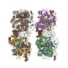
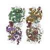
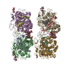
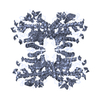
| Entry | Database: PDB / ID: 6ji1 | |||||||||
|---|---|---|---|---|---|---|---|---|---|---|
| Title | Tetrameric PepTSo2 incorporated in salipro nano particle | |||||||||
 Components Components | Proton:oligopeptide symporter POT family | |||||||||
 Keywords Keywords | MEMBRANE PROTEIN / Peptide transporter | |||||||||
| Function / homology |  Function and homology information Function and homology informationdipeptide transmembrane transport / tripeptide transmembrane transport / tripeptide transmembrane transporter activity / peptide:proton symporter activity / dipeptide transmembrane transporter activity / identical protein binding / plasma membrane Similarity search - Function | |||||||||
| Biological species |  Shewanella oneidensis MR-1 (bacteria) Shewanella oneidensis MR-1 (bacteria) | |||||||||
| Method | ELECTRON MICROSCOPY / single particle reconstruction / cryo EM / Resolution: 4.1 Å | |||||||||
 Authors Authors | Kawamoto, A. / Matoba, K. / Takagi, J. | |||||||||
| Funding support |  Japan, 2items Japan, 2items
| |||||||||
 Citation Citation | Journal: Acta Crystallogr F Struct Biol Commun / Year: 2019 Title: Structural basis for oligomerization of the prokaryotic peptide transporter PepT. Authors: Reina Nagamura / Masahiro Fukuda / Akihiro Kawamoto / Kyoko Matoba / Naoshi Dohmae / Ryuichiro Ishitani / Junichi Takagi / Osamu Nureki / Abstract: Proton-dependent oligopeptide transporters (POTs) belong to the major facilitator superfamily (MFS) and transport dipeptides and tripeptides from the extracellular environment into the target cell. ...Proton-dependent oligopeptide transporters (POTs) belong to the major facilitator superfamily (MFS) and transport dipeptides and tripeptides from the extracellular environment into the target cell. The human POTs PepT1 and PepT2 are also involved in the absorption of various orally ingested drugs. Previously reported structures revealed that the bacterial POTs possess 14 helices, of which H1-H6 and H7-H12 constitute the typical MFS fold and the residual two helices are involved in the cytoplasmic linker. PepT from Shewanella oneidensis is a unique POT which reportedly assembles as a 200 kDa tetramer. Although the previously reported structures suggested the importance of H12 for tetramer formation, the structural basis for the PepT-specific oligomerization remains unclear owing to the lack of a high-resolution tetrameric structure. In this study, the expression and purification conditions for tetrameric PepT were optimized. A single-particle cryo-EM analysis revealed the tetrameric structure of PepT incorporated into Salipro nanoparticles at 4.1 Å resolution. Furthermore, a combination of lipidic cubic phase (LCP) crystallization and an automated data-processing system for multiple microcrystals enabled crystal structures of PepT to be determined at resolutions of 3.5 and 3.9 Å. The present structures in a lipid bilayer revealed the detailed mechanism for the tetrameric assembly of PepT, in which a characteristic extracellular loop (ECL) interacts with two asparagine residues on H12 which were reported to be important for tetramerization and plays an essential role in oligomeric assembly. This study provides valuable insights into the oligomerization mechanism of this MFS-type transporter, which will further pave the way for understanding other oligomeric membrane proteins. | |||||||||
| History |
|
- Structure visualization
| Movie |
Movie viewer |
|---|---|
| Structure viewer | Molecule: MolmilJmol/JSmol |
- Downloads & links
-Download
| PDBx/mmCIF format | 6ji1.cif.gz | 310.6 KB | Display | PDBx/mmCIF format |
|---|---|---|---|---|
| PDB format | pdb6ji1.ent.gz | 252.6 KB | Display | PDB format |
| PDBx/mmJSON format | 6ji1.json.gz | Tree view | PDBx/mmJSON format | |
| Others |  Other downloads Other downloads |
-Validation report
| Arichive directory | https://data.pdbj.org/pub/pdb/validation_reports/ji/6ji1ftp://data.pdbj.org/pub/pdb/validation_reports/ji/6ji1 | HTTPS FTP |
|---|
-Related structure data
| Related structure data | 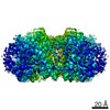 9832MC  6jkcC  6jkdC M: map data used to model this data C: citing same article ( |
|---|---|
| Similar structure data |


-Links
 PDBj
PDBj


- Assembly
| Deposited unit | 
|
|---|---|
| 1 |
|
-Components
| #1: Protein | Mass: 57626.559 Da / Num. of mol.: 4 Source method: isolated from a genetically manipulated source Source: (gene. exp.) Shewanella oneidensis MR-1 (bacteria) / Strain: MR-1 / Gene: SO_1277 / Production host: |
|---|
-Experimental details
-Experiment
| Experiment | Method: ELECTRON MICROSCOPY |
|---|---|
| EM experiment | Aggregation state: PARTICLE / 3D reconstruction method: single particle reconstruction |
- Sample preparation
| Component | Name: Tetrameric PepTSo2 with saposin A / Type: COMPLEX / Entity ID: all / Source: RECOMBINANT | |||||||||||||||
|---|---|---|---|---|---|---|---|---|---|---|---|---|---|---|---|---|
| Molecular weight | Experimental value: NO | |||||||||||||||
| Source (natural) | Organism: Shewanella oneidensis (bacteria) | |||||||||||||||
| Source (recombinant) | Organism: | |||||||||||||||
| Buffer solution | pH: 7.4 | |||||||||||||||
| Buffer component |
| |||||||||||||||
| Specimen | Conc.: 0.5 mg/ml / Embedding applied: NO / Shadowing applied: NO / Staining applied: NO / Vitrification applied: YES | |||||||||||||||
| Specimen support | Grid material: COPPER / Grid mesh size: 300 divisions/in. / Grid type: Quantifoil R1.2/1.3 | |||||||||||||||
| Vitrification | Instrument: FEI VITROBOT MARK IV / Cryogen name: ETHANE / Humidity: 100 % / Chamber temperature: 277 K / Details: blotted for 4.5 seconds before plunging |
- Electron microscopy imaging
| Experimental equipment |  Model: Talos Arctica / Image courtesy: FEI Company |
|---|---|
| Microscopy | Model: FEI TALOS ARCTICA |
| Electron gun | Electron source:  FIELD EMISSION GUN / Accelerating voltage: 200 kV / Illumination mode: FLOOD BEAM FIELD EMISSION GUN / Accelerating voltage: 200 kV / Illumination mode: FLOOD BEAM |
| Electron lens | Mode: BRIGHT FIELD / Nominal magnification: 96000 X / Nominal defocus max: 3000 nm / Nominal defocus min: 1000 nm / Cs: 2.7 mm / C2 aperture diameter: 50 µm / Alignment procedure: COMA FREE |
| Specimen holder | Cryogen: NITROGEN / Temperature (max): 79.55 K / Temperature (min): 79.55 K |
| Image recording | Electron dose: 82 e/Å2 / Detector mode: COUNTING / Film or detector model: FEI FALCON III (4k x 4k) / Num. of real images: 1609 |
| Image scans | Width: 4096 / Height: 4096 |
- Processing
| Software | Name: PHENIX / Version: (1.13_2998: phenix.real_space_refine) / Classification: refinement | ||||||||||||||||||||||||||||||||||||||||
|---|---|---|---|---|---|---|---|---|---|---|---|---|---|---|---|---|---|---|---|---|---|---|---|---|---|---|---|---|---|---|---|---|---|---|---|---|---|---|---|---|---|
| EM software |
| ||||||||||||||||||||||||||||||||||||||||
| CTF correction | Type: NONE | ||||||||||||||||||||||||||||||||||||||||
| Particle selection | Num. of particles selected: 288417 | ||||||||||||||||||||||||||||||||||||||||
| Symmetry | Point symmetry: C4 (4 fold cyclic) | ||||||||||||||||||||||||||||||||||||||||
| 3D reconstruction | Resolution: 4.1 Å / Resolution method: FSC 0.143 CUT-OFF / Num. of particles: 43172 / Algorithm: BACK PROJECTION / Symmetry type: POINT | ||||||||||||||||||||||||||||||||||||||||
| Atomic model building | Protocol: FLEXIBLE FIT / Space: REAL |
