 Movie
Movie Controller

[English] 日本語
 Yorodumi
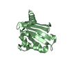
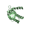
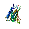
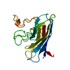
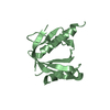
- PDB-6jhe: Crystal Structure of Bacillus subtilis SigW domain 4 in complexed... -
+ Open data

- Basic information
| Entry | Database: PDB / ID: 6jhe | ||||||
|---|---|---|---|---|---|---|---|
| Title | Crystal Structure of Bacillus subtilis SigW domain 4 in complexed with -35 element DNA | ||||||
 Components Components |
| ||||||
 Keywords Keywords | TRANSCRIPTION/DNA / protein-DNA complex / helix-turn-helix / TRANSCRIPTION-DNA complex | ||||||
| Function / homology |  Function and homology information Function and homology informationresponse to stress / sigma factor activity / DNA-templated transcription initiation / regulation of DNA-templated transcription / DNA binding Similarity search - Function | ||||||
| Biological species |  synthetic construct (others) | ||||||
| Method |  X-RAY DIFFRACTION / SYNCHROTRON / MOLECULAR REPLACEMENT / Resolution: 3.101 Å X-RAY DIFFRACTION / SYNCHROTRON / MOLECULAR REPLACEMENT / Resolution: 3.101 Å | ||||||
| Model details | Crystal Structure of Bacillus subtilis SigW domain 4 in complexed with -35 element DNA | ||||||
 Authors Authors | Kwon, E. / Devkota, S.R. / Pathak, D. / Dahal, P. / Kim, D.Y. | ||||||
 Citation Citation | Journal: Plos One / Year: 2019 Title: Structural analysis of the recognition of the -35 promoter element by SigW from Bacillus subtilis. Authors: Kwon, E. / Devkota, S.R. / Pathak, D. / Dahal, P. / Kim, D.Y. | ||||||
| History |
|
- Structure visualization
| Structure viewer | Molecule: MolmilJmol/JSmol |
|---|
- Downloads & links
-Download
| PDBx/mmCIF format | 6jhe.cif.gz | 61.6 KB | Display | PDBx/mmCIF format |
|---|---|---|---|---|
| PDB format | pdb6jhe.ent.gz | 43.3 KB | Display | PDB format |
| PDBx/mmJSON format | 6jhe.json.gz | Tree view | PDBx/mmJSON format | |
| Others |  Other downloads Other downloads |
-Validation report
| Summary document | 6jhe_validation.pdf.gz | 437.2 KB | Display | wwPDB validaton report |
|---|---|---|---|---|
| Full document | 6jhe_full_validation.pdf.gz | 441.1 KB | Display | |
| Data in XML | 6jhe_validation.xml.gz | 5.2 KB | Display | |
| Data in CIF | 6jhe_validation.cif.gz | 6.3 KB | Display | |
| Arichive directory | https://data.pdbj.org/pub/pdb/validation_reports/jh/6jheftp://data.pdbj.org/pub/pdb/validation_reports/jh/6jhe | HTTPS FTP |
-Related structure data
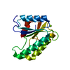
| Related structure data | 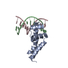 2h27S S: Starting model for refinement |
|---|---|
| Similar structure data |


-Links
 PDBj
PDBj


- Assembly
| Deposited unit | 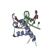
| ||||||||
|---|---|---|---|---|---|---|---|---|---|
| 1 |
| ||||||||
| Unit cell |
|
-Components
| #1: Protein | Mass: 7380.744 Da / Num. of mol.: 1 Source method: isolated from a genetically manipulated source Source: (gene. exp.) Strain: 168 / Gene: sigW, ybbL, BSU01730 / Plasmid: pETDuet-1 / Production host: |
|---|---|
| #2: DNA chain | Mass: 3323.196 Da / Num. of mol.: 1 / Source method: obtained synthetically / Source: (synth.) synthetic construct (others) |
| #3: DNA chain | Mass: 3381.248 Da / Num. of mol.: 1 / Source method: obtained synthetically / Source: (synth.) synthetic construct (others) |
-Experimental details
-Experiment
| Experiment | Method: X-RAY DIFFRACTION / Number of used crystals: 1 |
|---|
- Sample preparation
| Crystal | Density Matthews: 2.55 Å3/Da / Density % sol: 51.68 % |
|---|---|
| Crystal grow | Temperature: 293 K / Method: batch mode / pH: 6.5 / Details: PEG 1500, isopropanol, CaCl2 |
-Data collection
| Diffraction | Mean temperature: 100 K / Serial crystal experiment: N |
|---|---|
| Diffraction source | Source: SYNCHROTRON / Site: PAL/PLS  / Beamline: 7A (6B, 6C1) / Wavelength: 0.97933 Å / Beamline: 7A (6B, 6C1) / Wavelength: 0.97933 Å |
| Detector | Type: ADSC QUANTUM 270 / Detector: CCD / Date: Aug 5, 2017 |
| Radiation | Protocol: SINGLE WAVELENGTH / Monochromatic (M) / Laue (L): M / Scattering type: x-ray |
| Radiation wavelength | Wavelength: 0.97933 Å / Relative weight: 1 |
| Reflection | Resolution: 3→30 Å / Num. obs: 3013 / % possible obs: 99.4 % / Redundancy: 15.1 % / Biso Wilson estimate: 69.04 Å2 / Rmerge(I) obs: 0.104 / Net I/σ(I): 62.8 |
| Reflection shell | Resolution: 3→3.18 Å / Rmerge(I) obs: 0.524 / Num. unique obs: 468 / % possible all: 100 |
- Processing
| Software |
| ||||||||||||||||||||||||||||||||||||||||
|---|---|---|---|---|---|---|---|---|---|---|---|---|---|---|---|---|---|---|---|---|---|---|---|---|---|---|---|---|---|---|---|---|---|---|---|---|---|---|---|---|---|
| Refinement | Method to determine structure: MOLECULAR REPLACEMENT Starting model: 2H27 Resolution: 3.101→29.842 Å / SU ML: 0.28 / Cross valid method: THROUGHOUT / σ(F): 1.34 / Phase error: 18.36
| ||||||||||||||||||||||||||||||||||||||||
| Solvent computation | Shrinkage radii: 0.9 Å / VDW probe radii: 1.11 Å | ||||||||||||||||||||||||||||||||||||||||
| Displacement parameters | Biso max: 168 Å2 / Biso mean: 69 Å2 / Biso min: 44.16 Å2 | ||||||||||||||||||||||||||||||||||||||||
| Refinement step | Cycle: final / Resolution: 3.101→29.842 Å
| ||||||||||||||||||||||||||||||||||||||||
| LS refinement shell | Resolution: 3.101→3.318 Å / Rfactor Rfree error: 0
| ||||||||||||||||||||||||||||||||||||||||
| Refinement TLS params. | Method: refined / Origin x: -3.941 Å / Origin y: -20.7209 Å / Origin z: -0.3 Å
| ||||||||||||||||||||||||||||||||||||||||
| Refinement TLS group | Selection details: all |
