 Movie
Movie Controller

[English] 日本語
 Yorodumi
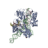
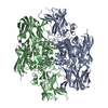
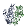
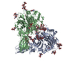
- PDB-6jfu: Crystal structure of Nme2Cas9 in complex with sgRNA and target DN... -
+ Open data

- Basic information
| Entry | Database: PDB / ID: 6jfu | ||||||
|---|---|---|---|---|---|---|---|
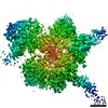
| Title | Crystal structure of Nme2Cas9 in complex with sgRNA and target DNA (AGGCCC PAM) | ||||||
 Components Components |
| ||||||
 Keywords Keywords | HYDROLASE/RNA/DNA / CRISPR-Cas9 / NmeCas9 / Nme2Cas9 / hydrolase / HYDROLASE-RNA-DNA complex | ||||||
| Function / homology | DNA / DNA (> 10) / RNA / RNA (> 10) / RNA (> 100) Function and homology information Function and homology information | ||||||
| Biological species |  Neisseria meningitidis (bacteria) Neisseria meningitidis (bacteria)synthetic construct (others) | ||||||
| Method |  X-RAY DIFFRACTION / SYNCHROTRON / MOLECULAR REPLACEMENT / Resolution: 3.2 Å X-RAY DIFFRACTION / SYNCHROTRON / MOLECULAR REPLACEMENT / Resolution: 3.2 Å | ||||||
 Authors Authors | Sun, W. / Yang, J. / Cheng, Z. / Liu, C. / Wang, K. / Huang, X. / Wang, Y. | ||||||
| Funding support |  China, 1items China, 1items
| ||||||
 Citation Citation | Journal: Mol.Cell / Year: 2019 Title: Structures of Neisseria meningitidis Cas9 Complexes in Catalytically Poised and Anti-CRISPR-Inhibited States. Authors: Sun, W. / Yang, J. / Cheng, Z. / Amrani, N. / Liu, C. / Wang, K. / Ibraheim, R. / Edraki, A. / Huang, X. / Wang, M. / Wang, J. / Liu, L. / Sheng, G. / Yang, Y. / Lou, J. / Sontheimer, E.J. / Wang, Y. | ||||||
| History |
|
- Structure visualization
| Structure viewer | Molecule: MolmilJmol/JSmol |
|---|
- Downloads & links
-Download
| PDBx/mmCIF format | 6jfu.cif.gz | 271.7 KB | Display | PDBx/mmCIF format |
|---|---|---|---|---|
| PDB format | pdb6jfu.ent.gz | 198.9 KB | Display | PDB format |
| PDBx/mmJSON format | 6jfu.json.gz | Tree view | PDBx/mmJSON format | |
| Others |  Other downloads Other downloads |
-Validation report
| Arichive directory | https://data.pdbj.org/pub/pdb/validation_reports/jf/6jfuftp://data.pdbj.org/pub/pdb/validation_reports/jf/6jfu | HTTPS FTP |
|---|
-Related structure data
| Related structure data |  6jdqC  6jdvSC  6je3C  6je4C  6je9C  6kc7C  6kc8C S: Starting model for refinement C: citing same article ( |
|---|---|
| Similar structure data |


-Links
 PDBj
PDBj


- Assembly
| Deposited unit | 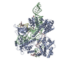
| ||||||||
|---|---|---|---|---|---|---|---|---|---|
| 1 |
| ||||||||
| Unit cell |
|
-Components
| #1: Protein | Mass: 124913.133 Da / Num. of mol.: 1 Source method: isolated from a genetically manipulated source Source: (gene. exp.) Neisseria meningitidis (bacteria) / Strain: DE10444 / Gene: cas9 / Production host: | ||||
|---|---|---|---|---|---|
| #2: RNA chain | Mass: 43059.352 Da / Num. of mol.: 1 / Source method: obtained synthetically / Source: (synth.) synthetic construct (others) | ||||
| #3: DNA chain | Mass: 10845.010 Da / Num. of mol.: 1 / Source method: obtained synthetically / Source: (synth.) synthetic construct (others) | ||||
| #4: DNA chain | Mass: 3358.211 Da / Num. of mol.: 1 / Source method: obtained synthetically / Source: (synth.) synthetic construct (others) | ||||
| #5: Chemical |   Mass: 62.068 Da / Num. of mol.: 2 / Source method: obtained synthetically / Formula: C2H6O2 Mass: 62.068 Da / Num. of mol.: 2 / Source method: obtained synthetically / Formula: C2H6O2Has protein modification | Y | Sequence details | Sequence of the protein has been deposited to NCBI with accession ID WP_002230835.1 | |
-Experimental details
-Experiment
| Experiment | Method: X-RAY DIFFRACTION / Number of used crystals: 1 |
|---|
- Sample preparation
| Crystal | Density Matthews: 3.16 Å3/Da / Density % sol: 61.08 % |
|---|---|
| Crystal grow | Temperature: 289 K / Method: evaporation / pH: 5.6 Details: 10% PEG 6000, 1.2M LiCl, 0.1M citrate acid pH 5.6, 4% 1,1,1,3,3,3-Hexafluoro-2-propanol |
-Data collection
| Diffraction | Mean temperature: 100 K / Serial crystal experiment: N |
|---|---|
| Diffraction source | Source: SYNCHROTRON / Site: SSRF / Beamline: BL17U1 / Wavelength: 0.97916 Å |
| Detector | Type: DECTRIS PILATUS3 S 6M / Detector: PIXEL / Date: Dec 6, 2018 |
| Radiation | Protocol: SINGLE WAVELENGTH / Monochromatic (M) / Laue (L): M / Scattering type: x-ray |
| Radiation wavelength | Wavelength: 0.97916 Å / Relative weight: 1 |
| Reflection | Resolution: 3.2→50 Å / Num. obs: 38356 / % possible obs: 99.7 % / Redundancy: 7.3 % / CC1/2: 1 / Rmerge(I) obs: 0.243 / Rpim(I) all: 0.093 / Rrim(I) all: 0.261 / Χ2: 0.956 / Net I/σ(I): 10.2 |
| Reflection shell | Resolution: 3.2→3.26 Å / Redundancy: 5.7 % / Rmerge(I) obs: 0.739 / Mean I/σ(I) obs: 2.8 / Num. unique obs: 1879 / CC1/2: 0.837 / Rpim(I) all: 0.323 / Rrim(I) all: 0.81 / Χ2: 0.908 / % possible all: 99.6 |
- Processing
| Software |
| ||||||||||||||||||||||||||||||||||||||||||||||||||||||||||||||||||||||||||||||||||||
|---|---|---|---|---|---|---|---|---|---|---|---|---|---|---|---|---|---|---|---|---|---|---|---|---|---|---|---|---|---|---|---|---|---|---|---|---|---|---|---|---|---|---|---|---|---|---|---|---|---|---|---|---|---|---|---|---|---|---|---|---|---|---|---|---|---|---|---|---|---|---|---|---|---|---|---|---|---|---|---|---|---|---|---|---|---|
| Refinement | Method to determine structure: MOLECULAR REPLACEMENT Starting model: 6JDV Resolution: 3.2→48.681 Å / SU ML: 0.39 / Cross valid method: FREE R-VALUE / σ(F): 1.39 / Phase error: 28.72
| ||||||||||||||||||||||||||||||||||||||||||||||||||||||||||||||||||||||||||||||||||||
| Solvent computation | Shrinkage radii: 0.9 Å / VDW probe radii: 1.11 Å | ||||||||||||||||||||||||||||||||||||||||||||||||||||||||||||||||||||||||||||||||||||
| Refinement step | Cycle: LAST / Resolution: 3.2→48.681 Å
| ||||||||||||||||||||||||||||||||||||||||||||||||||||||||||||||||||||||||||||||||||||
| Refine LS restraints |
| ||||||||||||||||||||||||||||||||||||||||||||||||||||||||||||||||||||||||||||||||||||
| LS refinement shell |
|
