 Movie
Movie Controller


+ Open data

- Basic information
| Entry | Database: PDB / ID: 1f13 | ||||||
|---|---|---|---|---|---|---|---|
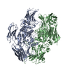
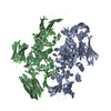
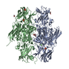
| Title | RECOMBINANT HUMAN CELLULAR COAGULATION FACTOR XIII | ||||||
 Components Components | CELLULAR COAGULATION FACTOR XIII ZYMOGEN | ||||||
 Keywords Keywords | COAGULATION FACTOR / COAGULATION / TRANSGLUTAMINASE / TRANSFERASE / ACYLTRANSFERASE / BLOOD COAGULATION | ||||||
| Function / homology |  Function and homology information Function and homology informationprotein-glutamine gamma-glutamyltransferase / peptide cross-linking / protein-glutamine gamma-glutamyltransferase activity / blood coagulation, fibrin clot formation / : / platelet alpha granule lumen / blood coagulation / Platelet degranulation / extracellular matrix / Interleukin-4 and Interleukin-13 signaling ...protein-glutamine gamma-glutamyltransferase / peptide cross-linking / protein-glutamine gamma-glutamyltransferase activity / blood coagulation, fibrin clot formation / : / platelet alpha granule lumen / blood coagulation / Platelet degranulation / extracellular matrix / Interleukin-4 and Interleukin-13 signaling / blood microparticle / : / extracellular region / metal ion binding Similarity search - Function | ||||||
| Biological species |  Homo sapiens (human) Homo sapiens (human) | ||||||
| Method |  X-RAY DIFFRACTION / SYNCHROTRON / MOLECULAR REPLACEMENT / Resolution: 2.1 Å X-RAY DIFFRACTION / SYNCHROTRON / MOLECULAR REPLACEMENT / Resolution: 2.1 Å | ||||||
 Authors Authors | Weiss, M.S. / Hilgenfeld, R. | ||||||
 Citation Citation | Journal: FEBS Lett. / Year: 1998 Title: Two non-proline cis peptide bonds may be important for factor XIII function. Authors: Weiss, M.S. / Metzner, H.J. / Hilgenfeld, R. #1: Journal: Thromb.Res. / Year: 1995Title: Structural Evidence that the Activation Peptide is not Released Upon Thrombin Cleavage of Factor Xiii Authors: Yee, V.C. / Pedersen, L.C. / Bishop, P.D. / Stenkamp, R.E. / Teller, D.C. #2: Journal: Proc.Natl.Acad.Sci.USA / Year: 1994Title: Three-Dimensional Structure of a Transglutaminase: Human Blood Coagulation Factor Xiii Authors: Yee, V.C. / Pedersen, L.C. / Le Trong, I. / Bishop, P.D. / Stenkamp, R.E. / Teller, D.C. #3: Journal: FEBS Lett. / Year: 1990Title: Crystallization of Blood Coagulation Factor Xiii by an Automated Procedure Authors: Hilgenfeld, R. / Liesum, A. / Storm, R. / Metzner, H.J. / Karges, H.E. | ||||||
| History |
|
- Structure visualization
| Structure viewer | Molecule: MolmilJmol/JSmol |
|---|
- Downloads & links
-Download
| PDBx/mmCIF format | 1f13.cif.gz | 300.2 KB | Display | PDBx/mmCIF format |
|---|---|---|---|---|
| PDB format | pdb1f13.ent.gz | 243.2 KB | Display | PDB format |
| PDBx/mmJSON format | 1f13.json.gz | Tree view | PDBx/mmJSON format | |
| Others |  Other downloads Other downloads |
-Validation report
| Arichive directory | https://data.pdbj.org/pub/pdb/validation_reports/f1/1f13ftp://data.pdbj.org/pub/pdb/validation_reports/f1/1f13 | HTTPS FTP |
|---|
-Related structure data
| Related structure data | 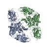 1ggtS S: Starting model for refinement |
|---|---|
| Similar structure data |


-Links
 PDBj
PDBj


- Assembly
| Deposited unit | 
| ||||||||
|---|---|---|---|---|---|---|---|---|---|
| 1 |
| ||||||||
| Unit cell |
| ||||||||
| Noncrystallographic symmetry (NCS) | NCS oper: (Code: given Matrix: (-0.395373, -0.917773, 0.037045), Vector: Details | THERE ARE TWO CHAINS IN THE ASYMMETRIC UNIT WITH CHAIN IDENTIFIERS A AND B. DISORDERED REGIONS INCLUDE RESIDUES 1 - 4, 37 - 38, 729 - 731 OF CHAIN A AND RESIDUES 1 - 5, 37 - 40, 729 - 731 OF CHAIN B. POORLY ORDERED REGIONS ARE RESIDUES 5 - 6 OF CHAIN A AND 6 OF CHAIN B, RESIDUES 35 - 36, 39, 510 - 516 OF CHAIN A AND 35 - 36, AND 510 - 516 OF CHAIN B. | |
-Components
| #1: Protein | Mass: 83233.922 Da / Num. of mol.: 2 / Fragment: A2-HOMODIMER Source method: isolated from a genetically manipulated source Source: (gene. exp.) Homo sapiens (human) / Organ: PLACENTA / Production host:  References: UniProt: P00488, protein-glutamine gamma-glutamyltransferase #2: Water | ChemComp-HOH / |  Mass: 18.015 Da / Num. of mol.: 487 / Source method: isolated from a natural source / Formula: H2O Mass: 18.015 Da / Num. of mol.: 487 / Source method: isolated from a natural source / Formula: H2O |
|---|
-Experimental details
-Experiment
| Experiment | Method: X-RAY DIFFRACTION / Number of used crystals: 5 |
|---|
- Sample preparation
| Crystal | Density Matthews: 2.86 Å3/Da / Density % sol: 57.1 % | ||||||||||||||||||||
|---|---|---|---|---|---|---|---|---|---|---|---|---|---|---|---|---|---|---|---|---|---|
| Crystal grow | pH: 6.2 Details: THE PROTEIN WAS CRYSTALLIZED FROM 1-2% PEG 6000, 100 MM MES, PH 6.2-6.4 PH range: 6.2-6.4 | ||||||||||||||||||||
| Crystal | *PLUS Density % sol: 57 % | ||||||||||||||||||||
| Crystal grow | *PLUS Method: vapor diffusion, hanging drop / PH range low: 6.4 / PH range high: 6.2 | ||||||||||||||||||||
| Components of the solutions | *PLUS
|
-Data collection
| Diffraction | Mean temperature: 293 K |
|---|---|
| Diffraction source | Source: SYNCHROTRON / Site: EMBL/DESY, HAMBURG  / Beamline: X11 / Wavelength: 0.912 / Beamline: X11 / Wavelength: 0.912 |
| Detector | Type: MARRESEARCH / Detector: IMAGE PLATE / Date: Sep 5, 1996 |
| Radiation | Monochromatic (M) / Laue (L): M / Scattering type: x-ray |
| Radiation wavelength | Wavelength: 0.912 Å / Relative weight: 1 |
| Reflection | Resolution: 2.1→99 Å / Num. obs: 89672 / % possible obs: 81.6 % / Observed criterion σ(I): 0 / Redundancy: 3.3 % / Rmerge(I) obs: 0.103 / Net I/σ(I): 10.7 |
| Reflection shell | Resolution: 2.1→2.18 Å / Redundancy: 1.9 % / Rmerge(I) obs: 0.3 / Mean I/σ(I) obs: 2.2 / % possible all: 40.2 |
| Reflection | *PLUS Num. measured all: 293385 |
- Processing
| Software |
| ||||||||||||||||||||||||||||||||||||||||||||||||||||||||||||
|---|---|---|---|---|---|---|---|---|---|---|---|---|---|---|---|---|---|---|---|---|---|---|---|---|---|---|---|---|---|---|---|---|---|---|---|---|---|---|---|---|---|---|---|---|---|---|---|---|---|---|---|---|---|---|---|---|---|---|---|---|---|
| Refinement | Method to determine structure: MOLECULAR REPLACEMENT Starting model: PDB ENTRY 1GGT Resolution: 2.1→40 Å / Data cutoff high absF: 10000000000 / Data cutoff low absF: 0 / Isotropic thermal model: RESTRAINED / Cross valid method: FREE R / σ(F): 0 / Details: BULK SOLVENT MODEL INCLUDED
| ||||||||||||||||||||||||||||||||||||||||||||||||||||||||||||
| Displacement parameters | Biso mean: 39.7 Å2 | ||||||||||||||||||||||||||||||||||||||||||||||||||||||||||||
| Refine analyze | Luzzati coordinate error obs: 0.25 Å / Luzzati d res low obs: 40 Å | ||||||||||||||||||||||||||||||||||||||||||||||||||||||||||||
| Refinement step | Cycle: LAST / Resolution: 2.1→40 Å
| ||||||||||||||||||||||||||||||||||||||||||||||||||||||||||||
| Refine LS restraints |
| ||||||||||||||||||||||||||||||||||||||||||||||||||||||||||||
| Refine LS restraints NCS | NCS model details: RESTRAINTS | ||||||||||||||||||||||||||||||||||||||||||||||||||||||||||||
| LS refinement shell | Resolution: 2.1→2.18 Å / Total num. of bins used: 10
| ||||||||||||||||||||||||||||||||||||||||||||||||||||||||||||
| Xplor file |
|
