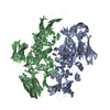
 Movie
Movie Controller


+ Open data

- Basic information
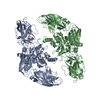
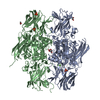
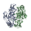
| Entry | Database: PDB / ID: 1qrk | |||||||||
|---|---|---|---|---|---|---|---|---|---|---|
| Title | HUMAN FACTOR XIII WITH STRONTIUM BOUND IN THE ION SITE | |||||||||
 Components Components | PROTEIN (COAGULATION FACTOR XIII) | |||||||||
 Keywords Keywords | TRANSFERASE / TRANSGLUTAMINASE / BLOOD COAGULATION / CALCIUM / STRONTIUM | |||||||||
| Function / homology |  Function and homology information Function and homology informationprotein-glutamine gamma-glutamyltransferase / protein-glutamine gamma-glutamyltransferase activity / peptide cross-linking / transferase complex / blood coagulation, fibrin clot formation / : / platelet alpha granule lumen / blood coagulation / Platelet degranulation / extracellular matrix ...protein-glutamine gamma-glutamyltransferase / protein-glutamine gamma-glutamyltransferase activity / peptide cross-linking / transferase complex / blood coagulation, fibrin clot formation / : / platelet alpha granule lumen / blood coagulation / Platelet degranulation / extracellular matrix / Interleukin-4 and Interleukin-13 signaling / blood microparticle / : / extracellular region / metal ion binding Similarity search - Function | |||||||||
| Biological species |  Homo sapiens (human) Homo sapiens (human) | |||||||||
| Method |  X-RAY DIFFRACTION / MOLECULAR REPLACEMENT / Resolution: 2.5 Å X-RAY DIFFRACTION / MOLECULAR REPLACEMENT / Resolution: 2.5 Å | |||||||||
 Authors Authors | Fox, B.A. / Yee, V.C. / Pederson, L.C. / Le Trong, I. / Bishop, P.D. / Stenkamp, R.E. / Teller, D.C. | |||||||||
 Citation Citation | Journal: J.Biol.Chem. / Year: 1999 Title: Identification of the calcium binding site and a novel ytterbium site in blood coagulation factor XIII by x-ray crystallography. Authors: Fox, B.A. / Yee, V.C. / Pedersen, L.C. / Le Trong, I. / Bishop, P.D. / Stenkamp, R.E. / Teller, D.C. | |||||||||
| History |
|
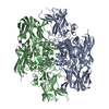
- Structure visualization
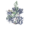
| Structure viewer | Molecule: MolmilJmol/JSmol |
|---|
- Downloads & links
-Download
| PDBx/mmCIF format | 1qrk.cif.gz | 289.9 KB | Display | PDBx/mmCIF format |
|---|---|---|---|---|
| PDB format | pdb1qrk.ent.gz | 233.5 KB | Display | PDB format |
| PDBx/mmJSON format | 1qrk.json.gz | Tree view | PDBx/mmJSON format | |
| Others |  Other downloads Other downloads |
-Validation report
| Arichive directory | https://data.pdbj.org/pub/pdb/validation_reports/qr/1qrkftp://data.pdbj.org/pub/pdb/validation_reports/qr/1qrk | HTTPS FTP |
|---|
-Related structure data
| Related structure data |  1gguC  1ggyC 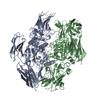 1fieS S: Starting model for refinement C: citing same article ( |
|---|---|
| Similar structure data |


-Links
 PDBj
PDBj


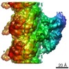
- Assembly
| Deposited unit | 
| ||||||||
|---|---|---|---|---|---|---|---|---|---|
| 1 |
| ||||||||
| Unit cell |
|
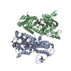
-Components
| #1: Protein | Mass: 83233.922 Da / Num. of mol.: 2 / Fragment: FULL LENGTH DIMER Source method: isolated from a genetically manipulated source Details: STRONTIUM BOUND IN THE ION SITE / Source: (gene. exp.) Homo sapiens (human) / Production host:  References: UniProt: P00488, protein-glutamine gamma-glutamyltransferase #2: Chemical |   Mass: 87.620 Da / Num. of mol.: 2 / Source method: obtained synthetically / Formula: Sr Mass: 87.620 Da / Num. of mol.: 2 / Source method: obtained synthetically / Formula: Sr#3: Water | ChemComp-HOH / |  Mass: 18.015 Da / Num. of mol.: 230 / Source method: isolated from a natural source / Formula: H2O Mass: 18.015 Da / Num. of mol.: 230 / Source method: isolated from a natural source / Formula: H2OSequence details | RESIDUES NOT VISIBLE IN ELECTRON DENSITY MAPS: CHAIN A 1-8, 31-44, 512-516, 728-731 CHAIN B 1-9, 36- ...RESIDUES NOT VISIBLE IN ELECTRON DENSITY MAPS: CHAIN A 1-8, 31-44, 512-516, 728-731 CHAIN B 1-9, 36-40, 508-515, 728-731 | |
|---|
-Experimental details
-Experiment
| Experiment | Method: X-RAY DIFFRACTION / Number of used crystals: 1 |
|---|
- Sample preparation
| Crystal | Density Matthews: 2.7 Å3/Da / Density % sol: 57 % | |||||||||||||||
|---|---|---|---|---|---|---|---|---|---|---|---|---|---|---|---|---|
| Crystal grow | Temperature: 298 K / Method: vapor diffusion, sitting drop / pH: 6.2 Details: 1,2-PROPANEDIOL (24%) AND NAKPO4 (100MM) PH 6.2, VAPOR DIFFUSION, SITTING DROP, temperature 25K | |||||||||||||||
| Crystal grow | *PLUS Method: unknown | |||||||||||||||
| Components of the solutions | *PLUS
|
-Data collection
| Diffraction | Mean temperature: 293 K |
|---|---|
| Diffraction source | Source: ROTATING ANODE / Type: RIGAKU RU200 / Wavelength: 1.5418 |
| Detector | Type: RIGAKU RAXIS IIC / Detector: IMAGE PLATE / Date: Jun 1, 1994 |
| Radiation | Protocol: SINGLE WAVELENGTH / Monochromatic (M) / Laue (L): M / Scattering type: x-ray |
| Radiation wavelength | Wavelength: 1.5418 Å / Relative weight: 1 |
| Reflection | Resolution: 2.5→10 Å / Num. obs: 50777 / % possible obs: 78.5 % / Observed criterion σ(F): 1 / Observed criterion σ(I): 2 / Biso Wilson estimate: 17.6 Å2 |
| Reflection shell | Resolution: 2.5→2.65 Å / % possible all: 55.1 |
| Reflection shell | *PLUS % possible obs: 51 % / Mean I/σ(I) obs: 2.3 |
- Processing
| Software |
| ||||||||||||||||||||||||||||||||||||||||||||||||||||||||||||||||||||||||||||||||
|---|---|---|---|---|---|---|---|---|---|---|---|---|---|---|---|---|---|---|---|---|---|---|---|---|---|---|---|---|---|---|---|---|---|---|---|---|---|---|---|---|---|---|---|---|---|---|---|---|---|---|---|---|---|---|---|---|---|---|---|---|---|---|---|---|---|---|---|---|---|---|---|---|---|---|---|---|---|---|---|---|---|
| Refinement | Method to determine structure: MOLECULAR REPLACEMENT Starting model: PDB ENTRY 1FIE Resolution: 2.5→10 Å / Rfactor Rfree error: 0.005 / Data cutoff high absF: 100000 / Data cutoff low absF: 0.1 / Isotropic thermal model: RESTRAINED / Cross valid method: THROUGHOUT / σ(F): 2 / σ(I): 1 / Stereochemistry target values: Engh & Huber
| ||||||||||||||||||||||||||||||||||||||||||||||||||||||||||||||||||||||||||||||||
| Displacement parameters | Biso mean: 33.8 Å2
| ||||||||||||||||||||||||||||||||||||||||||||||||||||||||||||||||||||||||||||||||
| Refine analyze |
| ||||||||||||||||||||||||||||||||||||||||||||||||||||||||||||||||||||||||||||||||
| Refinement step | Cycle: LAST / Resolution: 2.5→10 Å
| ||||||||||||||||||||||||||||||||||||||||||||||||||||||||||||||||||||||||||||||||
| Refine LS restraints |
| ||||||||||||||||||||||||||||||||||||||||||||||||||||||||||||||||||||||||||||||||
| LS refinement shell | Resolution: 2.5→2.65 Å / Rfactor Rfree error: 0.02 / Total num. of bins used: 6
| ||||||||||||||||||||||||||||||||||||||||||||||||||||||||||||||||||||||||||||||||
| Xplor file |
| ||||||||||||||||||||||||||||||||||||||||||||||||||||||||||||||||||||||||||||||||
| Software | *PLUS Name: X-PLOR / Version: 3.1 / Classification: refinement | ||||||||||||||||||||||||||||||||||||||||||||||||||||||||||||||||||||||||||||||||
| Refinement | *PLUS Lowest resolution: 10 Å / % reflection Rfree: 5 % | ||||||||||||||||||||||||||||||||||||||||||||||||||||||||||||||||||||||||||||||||
| Solvent computation | *PLUS | ||||||||||||||||||||||||||||||||||||||||||||||||||||||||||||||||||||||||||||||||
| Displacement parameters | *PLUS Biso mean: 33.8 Å2 | ||||||||||||||||||||||||||||||||||||||||||||||||||||||||||||||||||||||||||||||||
| Refine LS restraints | *PLUS
| ||||||||||||||||||||||||||||||||||||||||||||||||||||||||||||||||||||||||||||||||
| LS refinement shell | *PLUS Rfactor Rfree: 0.342 / % reflection Rfree: 4.7 % / Rfactor Rwork: 0.27 |
